Tuesday, December 11, 2012

2.0MP Handheld USB Digital Endoscope/Microscope with 8.2mm Tube Diameter

2.0MP Handheld USB Digital Endoscope/Microscope with 8.2mm Tube Diameter Review


2.0MP Handheld USB Digital Endoscope/Microscope with 8.2mm Tube Diameter Feature

  • Sensor: 2.0M pixels CMOS sensor. Still Image Resolution: 1600X1200 pixels (Format: BMP). Video Resolution: 1600x1200 pixels (Format: AVI, Frame Rate: 20FPS at VGA).
  • Focal Distance: 5mm - infinity (when Focal distance is infinity, the microscope cannot magnify, but can be used as a webcam). Magnification: 10X-200X (adjustable).
  • Illumination: 6 white LED lights built-in. PC Interface: USB 2.0. USB Cable Length: 165cm. Endoscope Tube Diameter: 8.2mm. Endoscope Tube Length: 160mm. Reflector Tube Diameter: 9.7mm.
  • Set of Accessories (Reflector Tube, Rubber Sleeve, Curette, and Plastic Sleeves): can be attached to the endoscope for different inspection purposes.
  • Compatibility: Windows 7(32 or 64 bit)/Vista/XP, MAC 10.5 or above (note: For Mac, please use Photo Booth or Facetime to open it. No software for Mac at this time). Manufacturer's Warranty: 1 year.

2.0MP Handheld USB Digital Endoscope/Microscope with 8.2mm Tube Diameter Overview

Connect digital endoscope to your computer with USB cable included, and inspect small details right on your computer screen! With 2.0M pixels CMOS sensor, up to 200x magnification, and six built-in LEDs for illumination, this product allows you to view images in high resolution, take standard photos, record videos, and use as a webcam. It is easy to take snapshot by hardware button or by software control, and the images can be saved, compared and printed via software. The endoscope comes with an adjustable aluminum alloy stand, and a set of accessories (reflector tube, rubber sleeve, curette, plastic sleeves) which can be attached to the endoscope for different inspection purposes. The endoscope is multi-functional. It can be used for inspecting hard-to-reach places such as the inside of engines, machines, tubes, pipes, etc. It is also ideal for a variety of applications including textile inspection, detailed repair, printing inspection, computer parts inspection, nature observation, reading aid, collectables examination (coins, antiques), and health (skin) examination. Package Includes: USB Digital Endoscope (1x), CD (1x, with User's Manual and software), Reflector Tube (1x), Rubber Sleeve (1x), Curette (1x), Small Plastic Sleeve (1x), Big Plastic Sleeve (1x), Adjustable Aluminum Alloy Stand (1x).

*** Product Information and Prices Stored: Dec 11, 2012 19:00:07

Thanks To : Finishing Products The Pneumatic Rackmount 19 inch waterproof digital camera canon

Monday, December 10, 2012

Digital Blue QX5 Digial Microscope

Digital Blue QX5 Digial Microscope Review


Digital Blue QX5 Digial Microscope Feature

  • Includes specimen jars, sample slide tweezers, eye dropper, slide clip, software CD-ROM, USB cable, microscope stand and microscope
  • Video playback 15 frames per second
  • Magnification 10X, 60X, 200X
  • Resolution VGA 640 x 480, twin super bright LED light source
  • Portable and easy to use in both stationary and "handheld" modes

Digital Blue QX5 Digial Microscope Overview

Get children to play the smart way with the Digital Blue series QX5 USB Computer Microscope. The QX5 magnifies anything from 10x all the way to 200x, and kids can use this microscope to view their favorite goodies - ants, bees wings, their school lunch...Well, they can use this to examine just about anything their little hearts desire! The QX5 is not only a microscope though, it also serves as a multi-function digital camera for taking digital still images and creating time-lapse movies. The QX5 also includes photo manipulation software that allows them to create and experiment with special effects and audio effects, which can all be inserted into the same images taken with the QX5. The QX5 microscope is a great toy for any child with an imagination, and will keep them happy for a long time to come! New Upgraded Model (from QX3) Now Featuring: Super-brite LED lighting for brighter, longer lasting top and bottom illumination, higher resolution 640x480 for more detailed images, video playback now three times faster at 15 frames per second, sample measurement tools built into the software.

*** Product Information and Prices Stored: Dec 10, 2012 12:30:03

Related : Picking Safety Products Measurement Guide Backup Mobile

Sunday, December 9, 2012

History And Uses Of optic Microscopes

Optical microscopes use descriptive light and a system of lenses to magnify small samples that are ordinarily un-seen to the bare eye. The optic microscope is the first, oldest and simples type of microscope (as opposed to the much more developed electronic microscope). The first optic microscopes were created in the 18th century. Due to it's compact sizes, simplicity and relatively low price, the optic microscope is very popular, and can be found in use in many areas of biology. optic microscopes mostly magnify objects for up to 1500 times.

The first optic microscopes were structured in a way that is called "the straightforward microscope". This buildings utilizes only one pair of lenses to create a magnified image of the sample. Today, the straightforward buildings is in use only in the magnifying glass, hand lens and the loupe.

Microscope

The more advances optic microscopes, and the ones that are favorite today, are what's called "compound optic microscopes". These microscopes use a system of many lenses, in order to "compound" and multiply the magnification, and therefore maximize it. The two main lens systems in an optic microscope are the objective lens (near the examined object), and the eyepiece lens (up near the eye of the scientist). Contemporary optic microscopes use complicated lenses both in the objective part as well as the eyepiece part.
The old optic microscopes also used a mirror to furnish illumination below the object. The Contemporary optic microscopes use a strong lamp to furnish constant and strong illumination.

So what are optic microscopes used for now a days?
The main uses of aggregate optic microscopes include:

The examining small pieces of material, or even a smear or a squash preparation. This is due to the fact that the optic microscope uses light to pass beneath the object and enter the lenses. That's why the item is good be half-transparent. In other uses the optic microscope may be used to observe metal samples, in order to study the metal's structure.
At low power, microscopes can be used to observe small living animals and plants. At high power, they can be used to observe bacteria.

It is important to note that the vast advancement in restorative fields and biology in general, is owed to a large extent, to the invention of the optic microscopes. For example, the way the blood flows in our body was not fully understood until the microscope made in inherent to observe small blood vessels behavior.

History And Uses Of optic Microscopes

Recommend : Material Handing Products The Pneumatic Pneumatic Plumbing Men Stainless Steel Watch

Friday, December 7, 2012

Light Microscopes

Light microscopes or moving field microscopes are the most basic form of microscopes. The traditional light microscope was invented way back in the 16th century. They are so called because they use light in the illustrated spectrum to illuminate the specimen. Light microscopes are extensively used in any place from high-tech laboratories to schools.

There are many types of light microscopes available from just to 00 or even more. The major types of light microscopes are stereo or dissecting, easy and composition microscopes. Stereo microscopes are the most advanced and use two light paths for three-dimensional (3D) viewing. They are very high-priced and are used in laboratories and medicinal practices. easy light microscopes are used only for small scale magnification and they have only one lens both as eyepiece and objective.

Microscope

Compound microscopes are the most extensively used light microscopes. There are mainly three grades of composition microscopes - student, benchtop and investigate light microscopes. Student microscopes are cheap, small and designed for moving field, dark field, and phase disagreement examinations. Benchtop microscopes are used for examination techniques. investigate microscopes are for investigate practices; they often include built-in computers and cameras.

The components of a tasteless composition microscope are: a mirror or light source for reflecting or producing light, a condenser with diaphragms and filters to adjust the intensity of the light, a stage for supporting the specimen which contains a central hole to pass the light on to the specimen, changeable objective lens for focusing the image for the eyepiece, fixed eyepiece or ocular for focusing the image for the eye, screws known as adjustments (coarse adjustment and fine adjustment) are used to bring the specimen in to focus, and a framework to which all these parts are attached. The power of the microscope depends on the power of the objective and eyepiece.

If you intend to buy a light microscope, go to specialist microscope suppliers, because only they can give you satisfying after-purchase services. The Internet also provides facts about some favorite suppliers. Often Internet sites offer you better deals and services.

Light Microscopes

Recommend : Finishing Products Picking Safety Products underwater canon digital camera

Thursday, December 6, 2012

Light Microscopes And Their Parts

The term "light microscope" is a general term that needs a bit of clarification. Just about all microscopes use a light source to illuminate the sample or specimen. This is why they are called light microscopes.

There are some types of light microscopes including straightforward (single lens) visual microscopes, blend (several lenses) microscopes, stereomicroscopes and digital microscopes. Light microscopes have varying levels of magnification available. With the irregularity of the stereomicroscope, these ordinarily have a magnification range somewhere between 20X and 1500X. The stereomicroscopes have a lower magnification range of between 20X and 200X due to the fact that they are used primarily to study the surfaces of larger specimens.

Microscope

In a nutshell, here is a list of the parts of a light microscope.

1. The base is just that: a base that the microscope stands on and allows it to be free standing.

2. The stage is where the sample or specimen slide is placed. There are two metal clips that hold the slide in place.

3. On the stage is a small opening in the center called an aperture. The sample is situated over the gap ,break for viewing and is held by the clip son the stage.

4. Below the stage is the light source. This is simply a small light bulb that shines upwards through the aperture, thus illuminating the sample. Some microscopes have a diaphragm in the stage that controls how much light is passed through the aperture. Note: Stereomicroscopes use two light sources to produce a 3D image to the viewer. The light source can be situated above the stage as in the case of digital microscopes. These microscopes are called inverted microscopes.

5. Directly above the stage is the nosepiece that contains the lenses used to magnify the sample. The nosepiece holds the lenses. The nosepiece rotates so the viewer can adopt the one they want to use.

6. The lenses, also called objectives, are held by the nosepiece and have dissimilar magnification powers, ordinarily from 2X to 15X or 20X.

7. The arm seems to associate the base to the upper parts of the microscope. It is used to carry the microscope.

8. The common adjustment knob is settled on the side of the microscope. It is used to focus the sample. This knob can move either the stage or the upper parts of the scope.

9. The fine adjustment knob is also settled on the side of the scope. This is used to fine-tune the focus after the common adjustment has brought the sample into view.

10. The body or tube holds the eyepiece(s) and connects it to the nosepiece lenses.

11. The eyepiece is what you look into. The eyepiece has a magnifying power of almost 10X.

So now you know all the parts of the microscope, you can intelligently tell your friends all about it. Knowing what the microscope's parts are helps the user to be more comfortable their first time out. It also helps if you need to order change parts later on.

Light Microscopes And Their Parts

Friends Link : Picking Safety Products Point of View Telescope waterproof camera panasonic

Wednesday, December 5, 2012

selecting The Best Microscope For Kids - Four Top Tips

A microscope is a great educational gift for a child. Here I'm going to tell you what to look for in a good microscope and where to get the best deal ready at the moment.

The microscope was invented in the late seventeenth century and for most of the time since then has been an expensive, pro instrument, often costing thousands of dollars. In new times though, the improvement of plastics and electronics has brought easy to use, remarkable microscopes within the reach of everyone.

Best Usb Microscope

Two sorts of microscope are ready for childrens' use: the customary turret type and the much newer electronic type that plugs into the Usb port of your Pc. I'll be writing about the Usb type in someone else article but here, if you'll excuse the pun, I'm going to focus on the turret type since these give children a much great idea of what true scientific research is like.
Look for a microscope with a metal body. These will be much heavier and therefore more garage when a child peers down the eyepiece. Small, lightweight microscopes move colse to all over the place when you try to look down them and are to be avoided at all costs. Try, if possible, to get a microscope with Led illumination. This type is fairly new but the Led type has the great benefit that the lights themselves create roughly no heat. It used to be a continual problem with filament lamp microscopes that the heat from the bulb would dry out specimens in next to no time. If kids are taking their time over what they're seeing at then they need lighting that won't ruin the specimens before they've beyond doubt seen what they are. Led lamps also have the added benefit of producing a whiter light than a filament bulb so the colors your kids will see will be all the more realistic. Make sure that the lenses are precision ground visual glass. visual glass is of higher density than, say, window glass, so, for a given magnification the lenses can be considerably thinner. This reduces what is a coarse problem in cheap lenses; it's called chromatic aberration and is the technical name for the colored rings colse to the edges of objects that you'll see when you look down your microscope. Make sure the microscope comes with a unblemished kit of accessories. Trying to dissect or manipulate specimens without the permissible tools just ruins them.

selecting The Best Microscope For Kids - Four Top Tips

Thanks To : Sun Solar cell Measurement Guide hard disk media player samsung batteries for cell phones

Tuesday, December 4, 2012

Konus 450x Konuspix Digital USB Microscope, Blue w/ 8 Blank Slides & Prep Accessories 5023

Konus 450x Konuspix Digital USB Microscope, Blue w/ 8 Blank Slides & Prep Accessories 5023 Review


$30.2

Konus 450x Konuspix Digital USB Microscope, Blue w/ 8 Blank Slides & Prep Accessories 5023 Feature

  • 8 Blank Slides
  • 4 prepared slides
  • Slide prep accessories

Konus 450x Konuspix Digital USB Microscope, Blue w/ 8 Blank Slides & Prep Accessories 5023 Overview

Konus 450x Konuspix Digital USB Microscope, Blue w/ 8 Blank Slides & Prep Accessories 5023

*** Product Information and Prices Stored: Dec 04, 2012 06:45:05
$89.79

Related : Picking Safety Products Top 10 Tablet PCs easy share cameras

Saturday, December 1, 2012

USB Flexible Inspection Camera Borescope Endoscope

USB Flexible Inspection Camera Borescope Endoscope Review


USB Flexible Inspection Camera Borescope Endoscope Feature

  • Sensor: 1/6" VGA CMOS. Image Resolution: 640 x 480 pixels (Format: JPEG).
  • Video Resolution: 640x480 pixels (Format: AVI, Frame Rate: up to 30FPS @ VGA). Focal Distance: 6cm - infinite. Lens: F#2.8 with 54° viewable angle.
  • Waterproof camera head & cable (handle is NOT waterproof) meet IP68 standard. PE covered cable with ABS handle.
  • Illumination: 2 white LED lights with adjustable brightness. USB Cable: 200cm (6ft) long with USB 1.1 interface.
  • Dimension: Flexible cable with handle: 88.0cm (2.9ft);
  • Flexible cable: 63.5cm (2.1ft); Flexible cable diameter: 8.5mm (0.35in); Camera head outer diameter: 14mm (0.55in).

USB Flexible Inspection Camera Borescope Endoscope Overview

By connecting the USB Flexible Inspection Camera to your PC via USB interface, it allows you to perform detailed visual inspection in hard-to-reach places. This inspection camera is powered by a USB connection to your PC, so no batteries are required. The 63.5cm (2.1ft) flexible cable can be bent into many shapes to fit into unreachable areas. With 1/6" VGA CMOS sensor and two built-in LEDs for illumination, this camera allows you to capture snapshots or videos with rich color throughout a large dynamic range. It is easy to take snapshot by hardware button or by software control, and the images can be saved, compared and printed via software. Waterproof camera withstands wet environments. In addition, the inspection camera comes with a magnetic accessory for picking-up nuts, bolts and screws that you may have dropped in inaccessible places. The small 14mm camera head is perfect for up-close visual inspections in small spaces. It is ideal for use in automotive maintenance, electrical repair, plumbing applications, HVAC/R and the visual inspection of otherwise inaccessible areas. Compatibility: Windows 7/Vista/XP. Package Includes: USB Inspection Camera, CD Software, Magnetic Pick Up Attachment, User's Manual, Plastic Carrying Case. Manufacturer's Warranty: 1 year. Note: All the Pictures, Technical Details, and Product Description above are provided by Oasis Scientific, Inc. We provide technical support to our customers.

*** Product Information and Prices Stored: Dec 01, 2012 07:45:04

Thanks To : The Fasteners Picking Safety Products hd lg led smart tv

Thursday, November 29, 2012

Check Out Dino-Lite Pro II AM411T Microscope for $329.00

Dino-Lite Pro II AM411T Microscope Review


Dino-Lite Pro II AM411T Microscope Feature

  • 1.3MP Resolution
  • USB2.0 Interface
  • 8 built-in LED Controllable with Software
  • Software supports:Windows 2000, XP, Vista; Mac OS 9,X
  • Built-in tap sensor for easy image capture

Dino-Lite Pro II AM411T Microscope Overview

The Dino-Lite Pro AM411T is a top selling Dino-Lite digital microscope for industrial inspection applications and hobbyists such as coin collectors and others. It boasts excellent color resolution in common with all Dino-Lite digital microscopes, a 1.3 megapixel camera, a digital "zoom in" feature up to 330x in VGA mode and a trigger pad, shooting mechanism on the end of the microscope. This combination makes for a highly versatile option for industrial inspection applications.In our opinion a key feature is the excellent color resolution which is unparalleled at this price point. It is better than most, more expensive digital microscopes and is achieved by use of a sophisticated jeweler's color balance. We are impressed!In addition, the Dino-Lite provides live imaging, USB 2.0 output interface and the upgraded DinoCapture 2.0 v1.2.9.x software allows for user-friendly photography including stills, video and even time-lapse. It can now import or export frames, save and reload notations and switch default output between BMP and JPG for greater flexibility and ease of use. Eight LED lights provide strong, clean lighting, as required and can be manually turned on and off with the software.

Related Products

Customer Reviews


*** Product Information and Prices Stored: Nov 29, 2012 18:00:04

Related : Best Fasteners Sun Solar cell philips hd blu ray player underwater canon digital camera

Wednesday, November 28, 2012

GSI High-Definition Scientific Digital LED Microscope, USB Video Connection to Computers/Laptops/Notebooks, Magnifies All Microscopic Articles 400x, Special Image Treatment, Includes Stand for Handsfree Use, for Hobby/Science/Education/Industrial- Black

GSI High-Definition Scientific Digital LED Microscope, USB Video Connection to Computers/Laptops/Notebooks, Magnifies All Microscopic Articles 400x, Special Image Treatment, Includes Stand for Handsfree Use, for Hobby/Science/Education/Industrial- Black Review


GSI High-Definition Scientific Digital LED Microscope, USB Video Connection to Computers/Laptops/Notebooks, Magnifies All Microscopic Articles 400x, Special Image Treatment, Includes Stand for Handsfree Use, for Hobby/Science/Education/Industrial- Black Feature

  • Microscope Lens (25x - 400x), 8 LED Light Source, Snap Function, 5x Digital Zoom
  • Color CMOS Image Censor, High Speed CPU, 1280x960 Resolution, 2M Pixels Lens
  • USB Compatible for Instant Video Viewing on Computer or Laptop Screen
  • Auto White Balance Control, Sharpness Control, Color Mangement, Brightness Adjustable
  • High Quality Image Enlarges Microscopic Articles on Skin, Cloth, Leather, Coins, Bills and Hair, Etc.

GSI High-Definition Scientific Digital LED Microscope, USB Video Connection to Computers/Laptops/Notebooks, Magnifies All Microscopic Articles 400x, Special Image Treatment, Includes Stand for Handsfree Use, for Hobby/Science/Education/Industrial- Black Overview

This season discover the Micro world with this newest innovation from GSI. This product was designed by our World-Class Micro experts to capture and display the tiniest miniscule objects in 400 times magnified viewing. Connect to your computer with USB cable included, simply point-and-Snap, and watch the world come alive in front of your eyes

*** Product Information and Prices Stored: Nov 28, 2012 08:30:04

Visit : The Fasteners Measurement Guide Fasteners Equipment Sonic Screwdriver Tool

Saturday, November 24, 2012

Celiac Disease Biopsy Explained: Part I Villous Atrophy

The determination of celiac disease is confirmed by a characteristic abnormal appearance of the small intestine under the microscope. Flattening of the normal finger like projections called villi accompanied by signs of inflammation is taken to indicate damage or injury from the storage protein gluten in wheat and similar proteins in barley and rye. The small intestine biopsy has became the gold proper for establishing the determination of Celiac disease or gluten sensitive enteropathy. Before 1960 gluten retirement followed by improvement and subsequent worsening upon rechallenge was the diagnostic criteria.

Early in the 1960's straight through the 1970's the small intestine was biopsied by having habitancy swallow a small metal capsule that was attached to a suction tube. This was used to suction up tissue into the capsule before guillotining off some tissue once the capsule was confirmed to be in the small intestine by x-ray. Now the tissue is obtained by upper endoscopy, the tube of a lighted video scope straight through the mouth under sedation to the small intestine, where biopsies are obtained with cupped forceps.

Microscope

Celiac disease biopsy: What does the pathologist look for under the microscope?

The small intestine normally has finger like projections called villi that give it a large face area or sense area for absorption. The villi result in a shag rug or terry cloth towel type appearance. Lining the face face of each villous are intestinal cells or enterocytes that secrete mucus and digest fluids, nutrients, minerals like iron, and vitamins like B12. On the face of the enterocytes are digestive enzymes like lactase that digest lactose or milk sugar. At the base of the villi are crypts or circular like collections of intestinal cells.

Celiac disease biopsy: What is villous atrophy?

Normally, villi are 3-5 times longer than the crypts are tall. However, intestinal injury can result in blunting, shortening (partial villous atrophy) or faultless loss of the villi and flattening (villous atrophy) of the intestinal surface. The shag rug will have bare spots or the terry cloth towel becomes like a tee shirt. The result is lack of absorption of nutrients and water resulting in weight loss, malnutrition, and diarrhea.

Celiac disease biopsy: What if the biopsy does not show atrophy or partial atrophy?

If the villi are at least 3 times as long as the crypts are tall then no flattening or blunting of the villi is gift and celiac disease becomes more difficult for the pathologist to diagnose without the history or blood test results. However, an increased estimate of Iel's (intra-epitheliel lymphocytes) in the setting of a distinct definite blood test for celiac, symptoms and especially if supported by presence of Dq2 and/or Dq8 gene pattern, is highly suggestive of celiac disease. The difficulty comes when the blood tests for the definite tests are negative or not elevated but only the "non-specific" blood tests (anti-gliadin or Aga and anti-reticulin antibodies) are elevated. Also, some habitancy with milder forms of celiac have no blood tests abnormal but have superior biopsy findings of celiac and are termed seronegative (blood test negative) celiacs.

Celiac disease biopsy: Can the biopsy be normal in celiac disease?

By definition, the biopsy has been determined the gold proper for diagnosing celiac. However, up-to-date studies have shown that the biopsy can be normal in some habitancy with celiac. How can this be? The pathologist reading the biopsy may explicate the biopsy as normal based on his or her bias about celiac disease, a failure to appreciate the importance of the presence of Iel's, or misuse of the older proper of >40 Iel's per 100. However, more importantly is the up-to-date recognition that normal appearing biopsies may not be normal. Electron microscopy has revealed ultra-structural abnormalities in apparent normal biopsies of habitancy confirmed to have celiac disease. Special stains, that consist of immune labeling of lymphocytes, have also confirmed increased numbers of distinct types of definite lymphocytes in the villi of intestinal biopsies of habitancy confirmed to have celiac. The bottom line is that a normal biopsy does not definitively exclude celiac disease or gluten sensitivity.

Celiac diasease biopsy: What are other inherent causes of biopsy changes that mimic celiac disease?

Cow's milk protein sensitive enteropathy (Cmse), viral or bacterial infections, medications (especially aspirin like arthritis medications e.g. Ibuprofen etc), autoimmune enteropathy, Helicobacter pylori infection (the stomach ulcer bacteria), Aids, tasteless variable immunodeficiency, and lymphoma of the intestine are all inherent causes of small intestine changes that may mimic celiac. However, if you have superior celiac type symptoms, a distinct celiac definite antibody (anti-endomysial antibody or tissue transglutaminase antibody) and a distinct response to a gluten free diet then celiac is the likely cause. The likelihood is supplementary increased if you carry one or both of the two major genes associated with celiac disease, Dq2 and/or Dq8. Normalization of celiac definite blood tests and the biopsy after a gluten free diet confirms the determination of celiac disease.

Celiac Disease Biopsy Explained: Part I Villous Atrophy

My Links : Pneumatics and Plumbing Good choice Sun Solar cell Pneumatic Plumbing light microscope parts hard disk media player

Thursday, November 22, 2012

AmScope 5x-10x-15x-30x Student Home School Digital Binocular Stereo Microscope with USB Imager Digital Camera

AmScope 5x-10x-15x-30x Student Home School Digital Binocular Stereo Microscope with USB Imager Digital Camera Review


$580.02

AmScope 5x-10x-15x-30x Student Home School Digital Binocular Stereo Microscope with USB Imager Digital Camera Feature

  • Stereo glass optical binocular offers four widefield magnification settings: 5X, 10X, 15X & 30X
  • Sturdy metal framework with pillar stand and built-in incident (top) lighting for examining 3D objects
  • 640x480 Pixel digital imager captures clear still images and saves them in BMP, TIFF, JPG, PICT, PTL or other format
  • User friendly software included for editing and processing microscope images, making measurement, recording video, and streams live videos on your PC screen
  • Compatible with Windows 2000/XP/Vista/7

AmScope 5x-10x-15x-30x Student Home School Digital Binocular Stereo Microscope with USB Imager Digital Camera Overview

This binocular stereo microscope comes with a 640x480 pixel USB digital camera. It has three magnification power settings, 5X, 10X, 15X & 30X, and an incident (top) lighting. This microscope offers high resolution and good depth within a broad field of view. It gives sharp clear stereo images. Its 45° inclined binocular head ensures an easy observation and rubber eyeguards provide further comfort. The digital camera captures microscope images and displays live video on your computer screen. With the user-friendly software included, compatible with Windows 2000/XP/Vista/7, you can edit microscope images, conduct measurement, record video, capture still images and save microscope images as a BMP, TIFF, JPG, PICT, PTL or other file. The camera dose not need any driver and the software is very easy to be installed on your computer. This microscopy system is a great instrument for many applications. It can be used in geology, forestry, archaeology, agriculture and biology. It is appropriate for educational purposes in hospitals and schools. It is great for the exploration of the world of surface details and fine structures of various materials like wood, metal, solder, semiconductor and stone. It is perfect for the examination of insects, plants, rocks, stamps, coins, jewel, soldered points, microelectronic components or virtually any three dimensional objects. This scope is an ideal tool for watchmaker, jeweler, engraver, dies-maker, philatelist, coin collector (not for viewing a whole coin), students, and etc. It comes with first-class quality and affordable price. It is brand new in factory-sealed box. The retail value of this microscope is 0.

*** Product Information and Prices Stored: Nov 23, 2012 00:00:04
$169.98

Thanks To : The Fasteners Picking Safety Products Measurement Guide blu ray player hd wifi signal booster for cell phone

Wednesday, November 21, 2012

7th Grade Science task Ideas Made uncomplicated and Fun

There are a whole of 7th grade science scheme ideas that a trainee can choose from to demonstrate concepts learned in class. A few such projects include:

o obtain water from a nearby stream or pond and view a drop of it under a microscope. Note what kinds of things you see and make drawings of them. Which are plants and which are animals? What is greater in your sample, plants or animals? What factors might influence the numbers of each that you find?

Microscope

o Draw a cell and label all of its parts. What function does each part of the cell serve? Are there differences in the middle of plant and animal cells? If do, what are they?

o Name the four different blood groups found in humans. What type is the rarest? The most common? How does the blood type of a person's parents influence the outcome of their own (i.e. If a person with type A blood marries person with type O, what type will any offspring have)?

o Draw a picture of a red blood call from a human and label all of its parts. Now draw a picture of the red blood cell from an animal, like a cow and label all of the parts. Are there any differences? If so, what part do you think these differences play in the function of the cell?

o Build a 3D model of a single-celled animal from cake. Use different tints of frosting to make the different structures inside the animal. Licorice laces can be cut up to describe the cilia. Before the cake is eaten, hand out a sheet that has the different parts labeled with call-outs, but not filled in. Allow people to view the cake and fill in the papers as to what each buildings is. Just before the cake is cut, put out a poster that shows what all is. Let the person who has the most right answers for these 7th grade science scheme ideas have the first piece of cake.

o Take a sample of cells from the inside of the mouth and view them under a microscope. Stain the material with a drop of food coloring. Communicate what you see. Make a drawing of any clear cells and label all of the structures present.

o Plankton is made up of several kinds of single cell plants and animals. Find out what they are and make drawings of several different kinds of each. What animals use plankton as a food source. What kinds of changes in the environment could turn plankton's survival in the water? Has plankton levels risen or fallen in recent years? Why?

o different bacteria exist all nearby us. Where are they found? What role do they play? In what cases should bacteria be encouraged to grow? In what cases should they be discouraged? Give an example of how or why they should be encouraged, and why they should be discouraged.

With a diminutive bit of particular understanding it is potential to find any whole of 7th grade science scheme ideas that can be turned into a science scheme for the inquisitive student.

7th Grade Science task Ideas Made uncomplicated and Fun

Thanks To : Finishing Products The Fasteners blu ray player hd wifi hard disk media player

Monday, November 19, 2012

What Are the Types of Microscopes?

An electron microscope can offer a man many separate types of advantages over other microscopes. If man has never used one of the many types of electron microscopes, then they are seriously missing out.

This type of microscope offers more clarity and a good magnification than most any other type of microscope on the market. In order to choose the best type of the many electron microscopes, a man must understand the various types that are ready for them to enjoy.

Best Usb Microscope

Three types of them exist. They are the transmission electron microscope, the scanning electron microscope, and the scanning-tunneling electron microscope. The original of all of these types of devices is the transmission electron.

The Tem, as it is commonly called, transmits electrons straight through a small slice of a specimen in order for the image to be seen. These types of devices are great for seeing inside the cells of beings on the planet. Much has been learned by using these microscopes for seeing all types of various cells.

Other types of the electron expedient are the scanning tunneling microscope and the scanning one. Both types of devices allow for a more in depth look than most other type of microscopes. The scanning microscope can absolutely allow scientists to view items in 3-D because of the excited atoms that are on a specimen.

The tunneling microscope absolutely creates a tunneling current in the middle of the electrons on a specimen and the microscopes tip to make the image. These are both very high tech microscopes that can be used to see farther into a specimen than previously conception possible.

An electron one can give a much more in depth view. Population once only had an idea of what cells and other indiscernible items looked like. Thanks to the invention of the electron microscopes and the various types that are available, a man can see these items in a three dimensional view that can make learning these items better. Microscopes are the wave of the future in today's current high tech world.

What Are the Types of Microscopes?

Related : Fasteners Equipment Measurement Guide light microscope magnification

Saturday, November 17, 2012

What is Upper Endoscopy and Why Is Small Intestine Biopsy Recommended for Celiac Disease?

When undergoing an appraisal for possible celiac disease or gluten sensitive enteropathy doctors commonly recommend an upper endoscopy and small intestine biopsy. What that may mean or why it is recommended may not be clear to habitancy who are facing the decision to endure the procedure themselves or to field their child to the exam.

Endoscopy in celiac: What is it and how is it done?

Microscope

The healing name for upper endoscopy is esophagogastroduodenoscopy or Egd for short. The endoscope is a thin flexible tube about the diameter of a fat pencil that has a video chip in the end and channels for flushing of water, suctioning of secretions and tube of instruments. It has dials that allow the tube to be turned up/down and right and left at the tip permitting it to be passed straight through the mouth, down the esophagus or feeding tube, into the stomach and then into the first part of the small intestine the duodenum, hence the name Egd.

Endoscopy in celiac: Do you feel it or remember it?

People undergoing the exam in the U.S. Typically are sedated with a medication. Medications similar to valium with good amnesia and relaxing supervene called midazolam or versed combined with a narcotic like meperidine (demerol) or fentanyl are generally used. More recently a very short acting intravenous sedative, propofol (diprovan), may be administered for deep sedation or an intravenous form of general anesthesia. Occasionally, commonly in very young children or habitancy with severe lung problems, general anesthesia is required. The exam is commonly not felt or remembered because of the medications.

Endoscopy in celiac: What is examined in celiac and how well can the lining be seen?

Celiac disease affects the upper measure of the small intestine, in the two sections known as the duodenum and jejunum. The exam of the small intestine is commonly exiguous to the first section termed the duodenum though occasionally the second section known as the jejunum may be reached especially when a longer endoscope is used. The resolution of video images are very high with the latest endoscopes and also may have a magnification and color contrast mode to detect very subtle signs of damage of the small intestine.

Endoscopy in celiac: What are the typical findings?

The characteristic appearance of the face of the small intestine in celiac disease consist of superficial ulcerations that are generally linear, flattening of the folds, notching or scalloping of the folds and a mosaic like pattern. However, the face may appear general and only under exiguous exam of samples will the lining show signs of gluten caused injury.

Endoscopy in celiac: What are biopsies?

Samples of small intestine are obtained with biopsy forceps that consist of tiny jaws with cups that permit pinching off samples of the intestinal lining. This is painless and very safe. The samples are sent to a determination lab in a preservative solution, processed, mounted on a microscope slide, and stained for exam under the microscope by a pathologist. Small intestine injury from gluten may be patchy, therefore, some samples are recommended. A minimum of 4 pieces and preferably 8-12 samples should be obtained to avoid missing exiguous signs of celiac disease.

Endoscopy in celiac: What does the pathologist look for on the slides?

The pathologist examines the slide for evidence of damage or injury characteristic of gluten sensitivity. Occasionally extra stains are required to see signs of irritation known as inflammation characterized by an increased estimate of a type of immune active white blood cells called lymphocytes. In early celiac and gluten sensitivity without celiac disease the biopsy may be general and the determination cannot be established by the biopsy.

Endoscopy in celiac: Summary.

The procedure of endoscopy is safe, painless, and very helpful for establishing the determination of celiac disease while excluding other upper intestinal disorders. The main drawback of endoscopy is that nearly everyone must have sedation to tolerate the exam and it can be high-priced if not fully covered by insurance. Sometimes, celiac disease is diagnosed by endoscopic biopsy in habitancy who whether have general blood tests or as an incidental seeing in those undergoing endoscopy for other reasons. Fear or blurring about endoscopy should not preclude anyone who is suspected of having celiac or gluten sensitivity from undergoing endoscopy. Additional information about celiac disease and other digestive diseases are available at http://www.thefooddoc.com, the premier website under amelioration by "the food doc", Dr. Scot Lewey, a practicing stomach and intestinal expert (gastroenterologist).

Copyright 2006, The Food Doc, Llc All ownership Reserved. Http://www.thefooddoc.com

What is Upper Endoscopy and Why Is Small Intestine Biopsy Recommended for Celiac Disease?

Tags : Measurement Guide Sonic Screwdriver Tool led smart tv sale

Friday, November 16, 2012

Check Out Celestron LCD Deluxe Digital Microscope (44345) for $216.93


Celestron LCD Deluxe Digital Microscope (44345) Overview

Celestron has developed the first digital LCD microscope with a 3.5" touch screen panel that is also rotatable 180 Degree for sharing the view on the screen. Also comes with a built-in digital camera, so you can take snapshots or video using the built-in flash memory or use an optional SD Card and then later upload your images to your computer.

Celestron LCD Deluxe Digital Microscope (44345) Review


Celestron LCD Deluxe Digital Microscope (44345) Feature

  • 3.5" TFT Touch Screen Panel - rotatable 180 Degree horizontally
  • Built-in Digital Camera - 2mp CMOS Sensor
  • Image Capture Capability - Snapshots and Video
  • Quadruple Nosepiece with 4x/10x/20x/40x Objective Lenses
  • Dual Focus (Coarse and Fine)

Related Products

Customer Reviews


*** Product Information and Prices Stored: Nov 16, 2012 13:15:06

Friends Link : Picking Safety Products inexpensive cell phone plans

Wednesday, November 14, 2012

Educational Microscopes

Educational microscopes play a essential role in today's education and research. As a great education tool, these microscopes help the students to view magnified images of all from plant cell walls to butterfly wings. Educational microscopes offer maximum flexibility and accuracy for viewing images.

As there are separate types of educational microscopes, the one chosen must best suit the student's requirement. A low-power microscope - also called stereo or dissecting microscope - is used to view larger things like stamps, coins, and sand grains. Mostly, it is supplied with 10X and 20X eye pieces. As it is easy to focus, this type of microscope is especially great for younger children. There are three models to pick from - particular power, two power, and zoom type stereo microscope.

Best Usb Microscope

High-power microscopes can be used to view itsybitsy objects like bacteria, plant cell, and blood samples. Their typical magnification is 40X, 100X, and 400X. High-power microscopes consist of biological microscopes, combination microscopes, and metallurgical microscopes. Educational combination microscopes are used for viewing the itsybitsy world. Their magnification can range up to 1000X. Tour Lab, MicroQuest, Explorer Ii, Observer Iii, and Observer Iv Binocular Microscope are some favorite models.

Digital microscopes are also greatly used for education purposes. These microscopes will help the students to view large images on a computer monitor, through a Usb connection. Thus, they allow the angle of a itsybitsy image to a large audience. Moreover, digital microscopes help the students to save and print the images.

Educational microscopes are equipped with triple or quadruple nosepiece, coaxial tasteless for spoton focusing, and reflecting mirror. A condenser with magnification indication is also offered. To get optimum resolution, the diaphragm of the condenser is provided with position guide markings for separate objectives. Additional features such as illuminator and stopper for slide security are also available.

Most educational microscopes are cordless and rechargeable. Some models are provided with a digital camera. Software for image capturing is also included. Apart from these, some microscope clubs offer Additional accessories such as dust cover, immersion oil, and prepared glass slides.

Educational Microscopes

Visit : Material Handing Products Men Eco Drive Watch kodack easy share

Monday, November 12, 2012

Carson Z-Pix 34-168x Digital Zoom Microscope


Carson Z-Pix 34-168x Digital Zoom Microscope


Carson Z-Pix 34-168x Digital Zoom Microscope The Carson Optical zPix is a powerful digital zoom microscope that displays a magnified image right on your computer screen. Impressive 34-to-168X magnification allows you to see details of ordinary objects you never knew existed, and you can additionally capture images with an onboard 1.3 megapixel digital camera.

Specifications

  • 34-to-168X magnification
  • Measures 1.8 by 4.1 by 1.7 inches (W x H x D)
  • 8.8-ounce weight
  • Compatible with Windows 98 SE, 2000, ME, XP, and Vista; Mac OS X 10.4 or later
  • Requires USB 2.0 port
  • Includes specimen base, three blank sides, one prepared slide, forceps, eyedropper, USB cable, and CD ROM software

About Carson Optical
Carson is a leading supplier of consumer optics products for major catalog companies, nationwide retailers, and direct to consumers. The company's product line covers categories from recreation, sports, hobby, education, and crafts with products ranging from high-end binoculars to low-cost magnifiers.

Recommend : Material Handing Products Measurement Guide Measurement Guide Point of View Telescope waterproof nikon digital camera

Saturday, November 10, 2012

Check Out Koolertron(TM) 5MP 8-LED USB Digital Microscope endoscope 5.0 Mega Pixels Magnifier Camera for $55.99


Koolertron(TM) 5MP 8-LED USB Digital Microscope endoscope 5.0 Mega Pixels Magnifier Camera Overview

Description :

USB Digital Microscope is a good quality to use in Inspection dissection/examination, Plant dissection/examination, Skin examination, Scal examination, Textile Inspection, Jewelry Inspection, Collections/Coin Inspection, Printing Inspection, PCB or PCBA Inspection and so on.
The microscope is made up of high-quality IC and electronic components,with clear image quality, low power consumption, high-resolution,applies to small objects to be larger? Photo and video production, and it is the best choice of small objects recognization.


Product Features:

Image sensor: 5Mega Pixels (Must Installion CD Drive)
Video Capture Resolution: 160x120, 320x240, 640x480, 1280x1024
Still Image Capture Resolution: 160x120, 320x240, 640x480, 1280x1024
Exposure: Auto
Photo format: JPEG
Light source: 8 LED
PC interface: Mini USB1.1&2.0
Power source: 5V DC from USB port
Gray scale: Level 8
Sleep current: 1 mA
Work current: 180 mA
Save temperature: -20°C to +60°C
Work temperature: 10°C to +40°C
Magnification Ratio: 20x to 200x
Operation system: Windows XP (32-bit system) /Vista (32-bit system) / Win7 (32-bit system)
?

Koolertron(TM) 5MP 8-LED USB Digital Microscope endoscope 5.0 Mega Pixels Magnifier Camera Review


Koolertron(TM) 5MP 8-LED USB Digital Microscope endoscope 5.0 Mega Pixels Magnifier Camera Feature

  • Video Capture Resolution: 160x120, 320x240, 640x480, 1280x1024
  • Still Image Capture Resolution: 160x120, 320x240, 640x480, 1280x1024
  • Exposure: Auto
  • Photo format: JPEG
  • Light source: 8 LED

Related Products

Customer Reviews


*** Product Information and Prices Stored: Nov 10, 2012 18:45:03

See Also : Pneumatics and Plumbing Good choice canon easy share camera

Friday, November 9, 2012

2.4Gz Wireless Endoscope available From China constructor Kasyn

Wireless EndoscopeA real novelty - 2.4Gz Scope-pro Wireless digital endoscope available already made in China from Kasyn.

The wireless endoscope is dissimilar from other general Usb digital microscopes. A general Usb digital endoscope will come with a Usb cable so that it can be associated with the Pc. But this Digital Microscope Endoscope is 2.4Gz wireless with a Usb dongle. The best efficient length is 10 meters. Kasyn suggests you try this item within this distance.

Best Usb Microscope

In order to see more clearly via this new endoscope, the make prepare a series of clear pictures "focus convert knob" tp to adjust the focus. Magnification is up to 200x Mp. There is no any tiny small traces you will miss via this professional scope-pro Wireless Digital Microscope.

Specification:

Working platforms: Windows 7, Vista, Xp, 2000, Mac, Linux

Magnification: up to 200x

Photograph: Taken by the software and touch button

Videogaphy: High compression

Light source: Led illumination

Video with ultra-long time-11hours long; 3.6Gb of memory video

Focusing: Manual-focus

Max Size: 11mm*126mm

Pixels: 1.3-5m

Display speed: 15-30fps

Interface: 2.4G wireless

Various Probes available, which can be more convenience and clear to search for dissimilar part of body, such as (air,Throat, Ears etc). It is certainly good machine for body beauty, home condition care.

Application: Golf procedure (to search for the grass health)

Workshops

Laboratories

Industrial inspection

Science education

Forensics

Printing

Textile

Printed circuit board (Pcb) inspection

Jewelers

Hobbyists

Tinkerers

Crime scene investigation

Medical

Serial number identification

Quality control

Parts assembly

Find a trustworthy victualer As a unique tech, you can only purchase this model via Kasyn International website. Kasyn dealer offers perfect technical hold also. They seems contribute a series of dissimilar endoscopes as well.

2.4Gz Wireless Endoscope available From China constructor Kasyn

Thanks To : Finishing Products Pneumatics and Plumbing Good choice Sun Solar cell high definition media player wd hd player

Thursday, November 8, 2012

The History of the Microscope

The first microscope was created hundreds of years ago. In the passing centuries, microscopes evolved into powerful, accurate tools that allow scientists to view tiny objects at a level of information that seems unreal. There are a wide array of available microscopes, from the aggregate microscopes commonly found in high school science classrooms to great scanning tunneling and electron microscopes used by Nobel Prize winners.

Most historians agree that two Dutchman made the first microscope in 1590. Zaccharias Janssen and his son Hans were two eyeglass makers who experimented with putting many lenses together in a tube. They found that objects under the tube were greatly enlarged. Over the next hundred years, scientists Robert Hooke, Anton van Leeuwenhoek, and others added refined the work of the Janssens and used microscopes to examine insects, blood, and other items. Scientists have continued microscopes into the gift day. Now, microscopes can show tiny particles that are unseen by the naked eye in extremely exact detail.

Microscope

Microscopes operate on several principles. Most coarse microscopes have two distinct lenses. Viewers look through the ocular lens, also known as the eyepiece. There is an additional one lens, called the objective lens at the end of the ocular lens. The objective lens is a sphere shaped lens located above the stage of the microscope. citizen place the object they want to examine on the stage and can adjust the lenses to bring the object into focus. Most microscopes have an adjustment knob for coarse focus and one for fine focus. Many microscopes have several objective lenses with distinct strengths for users to choose from. The lenses are arranged on a circular platform that can be rotated to have the distinct lenses put into place under the ocular lens. Microscopes also need a light source of some kind underneath the stage. Most commercial microscopes have a light bulb, but many high-end microscopes use lasers or electrons for illumination.

Microscopes have been used to make countless vital scientific discoveries. They are invaluable tools used in a range of scientific fields that enable researchers to make discoveries that would be impossible with the naked eye.

The History of the Microscope

Recommend : Fasteners Equipment cell phone covers samsung phones

Sunday, November 4, 2012

Firefly GT800 Handheld USB Digital Microscope with Measurement Functions

Firefly GT800 Handheld USB Digital Microscope with Measurement Functions Review


Firefly GT800 Handheld USB Digital Microscope with Measurement Functions Feature

  • Sensor Resolution: 2.0M pixels. Still Image Resolution: 1600x1200 pixels (Format: BMP). Video Resolution: 1600x1200 pixels (Formats: YUY2, AVI, Frame Rate: 30FPS at 640x480 pixels).
  • Magnification: 15x - 230x (Native Optical), 15x - 700x (Digital). Lens Assembly: Dual Lenses, 3-Layer Glass, 650nm cutoff.
  • Lighting: 8 Ultra-Bright LEDs with fully adjustable brightness. Interface: USB2.0 (USB Cable Length: 180cm). Built-in snapshot button. Rugged industrialized construction.
  • Software: Scalable Window, Zoom, Freeze, Resolution, Rotate, Flip Region of Interest (ROI), Real time measurements, Automatic/Manual white balance.
  • Safety Compliance: FCC, CE and RoHS compliant. Manufacturer's Warranty: 1 year.

Firefly GT800 Handheld USB Digital Microscope with Measurement Functions Overview

Firefly GT800 is one of the industry's most advanced high precision handheld digital microscopes, providing up to 230x native optical magnification and up to 700x digital magnification. Equipped with multi-layer glass lenses rather than plastic alternatives, this microscope delivers superb focal depth and crystal clear image quality. GT800 is controlled directly with the FireflyPro professional image processing software which is bundled with the microscope. This state-of-the-art software allows users to capture, store, recall, view, manipulate, and measure images and videos in real time. It is easy to take snapshot by hardware button or by software control. GT800 is ideal for a variety of applications including textile inspection, detailed repair, printing inspection, computer parts inspection, living creature examination, reading aid, and collectables examination (coins, stamps).Compatibility: Windows 7/Vista/XP. Package Includes: Microscope (1x), Stand (1x), USB Cable (1x), Software Application CD (1x User's Manual and software), Calibration Tool (1x), Velvet Carrying Case (1x).

*** Product Information and Prices Stored: Nov 04, 2012 10:45:03

Related : The Pneumatic Picking Safety Products Wall mount TV Choosing The Correct Tripods

Saturday, November 3, 2012

Polarizing Microscope and Applications

A polarizing microscope is a special kind of microscope that utilizes two polarizing lens to acquire positive optic data from the specimen. The polarizing microscope is used extensively in the field of optic mineralogy which supports such applications as geology, asbestos testing, and forensic science. Often those working in different fields will sometimes refer to the polarizing microscope by different names such as geology microscope, petrographic microscope, pol microscope, and Plm (polarized light microscope.)

The key unlikeness in the middle of the polarizing microscope and a proper aggregate microscope is the expanding of a fixed polarizer in the middle of the light source and the specimen and the expanding of an adjustable polarizer in the middle of the objective and the eyepieces. The 2nd polarizer is called the "analyzer" and normally can insert in and out on a rotating piece in the neck of the microscope. Other common accessories consist of a rotating stage and insertable retardation plates made from gypsum or quartz.

Microscope

With these additional elements, the this microscope can acquire optic data from a specimen that no other microscope can. The key optic data available includes refractive index, birefringence, sign of elongation, pleochroism, and angle of extinction, all of which supply clues to the crystallography of the material that is being investigated.

The first uses of these kinds of microscopes over one hundred years ago was the identification of minerals in geology. In addition, the most common form of lab analysis to test for asbestos is performed with a polarizing microscope. Because of the their ability to supply optic data, these kind of microscopes are very commonly used in forensic science where the identification of unknown materials is a routine part of the job. Some of the first evidence disputing the claimed age of the so-called "Shroud of Turin" was carefully by analysis of pigments using a polarized light microscope.

The polarizing microscope is a very versatile and mighty instrument in the identification of materials. It is a key tool in some scientific fields, and can sometimes be the best choice over more expensive technologies. For example, in routine asbestos analysis the polarizing microscope was carefully to be more correct and much more cost effective than the other high-tech options that were first investigated, such as x-ray diffraction and scanning electron microscopy. It is a mighty tool with many applications.

Nikon's MicroscopyU has quite a bit of data on how the polarizers and the science work to supply analytical information: http://www.microscopyu.com/articles/polarized/polarizedintro.html

Polarizing Microscope and Applications

Friends Link : Material Handing Products Picking Safety Products Pneumatics and Plumbing Good choice the compound microscope light microscope images

Friday, November 2, 2012

overview of the modern combination Light Microscope

The blend light microscope is one of the most commonly used types of microscope. Among its many uses is the science classroom, where they are an affordable and easy to use instrument for learning biology and chemistry. The instrument traces back generations, but recently has evolved into a very modern instrument.

Only a few decades ago, you could find the microscope in just two basic types. There was the monocular blend microscope and the binocular compound. That is, the blend light microscope came with either one or two viewing units. With the monocular compound, there was a single eyepiece and so you looked at your specimen with just one eye. This is still what many people envision when they hear the word "microscope." Then there's the binocular model which, naturally, resembles a pair of binoculars. That's because there are a pair of viewing units, one for each eye. Today, there are few if any monocular blend microscopes made. The new proper is the binocular.

Microscope

However, even more up-to-date times have introduced to us a newer standard. This is the trinocular blend microscope. The "tri" signifies a third viewing port. This makes it a not-as-common spin-off of the binocular compound.

The function of the trinocular microscope is a bit different, since most people only have two eyes. The third eyepiece, then, is for use by a second person. It comes in handy in a concentrate of situations. First, if two people need to consult together over the same specimen, they are able to look at it at the same time. A second and more common use of the trinocular is to allow a science teacher to see the same thing that his pupil sees under the microscope. A third good use for it is to take pictures of the specimen with a camera placed over the third eyepiece. Each of the microscope parts functions in harmony, allowing the camera to see what the person's eye sees.

The blend light microscope, as recommend earlier, is superior due to its pair of optical parts. Let's discuss these and how each of them is used.

1. First of these parts are the objective lenses on the scope. These lenses sit right above the specimen, collecting the light emitted by the specimen. The typical blend microscope has 3, 4 or 5 objective lenses. Each of these will offer a slightly dissimilar level of magnification. These objective lenses are held together by the nose piece. As this nose piece is notated, the magnification is altered by a dissimilar objective lens.

2) The eyepiece is the second major part. This is the part that your eye looks directly straight through to see the specimen. It's a cylinder shape and contains a lens. The eyepiece is also called the microscope ocular.

As these two major parts work together, there are is a huge range in magnification available. In fact, if it fits on the microscope, there is practically legitimately a magnification level that will bring it to life for you. It's virtually unlimited in the viewing inherent it offers you.

The blend scope has some other foremost parts, too, like the condenser. The condenser is one of the more brittle but foremost parts. There are other foremost parts to the microscope, such as the condenser. A diagram of the parts condenser is helpful to make sure it stays in good condition. Do a Google or Yahoo search for more information.

Costs for your microscope might vary, but ordinarily they will run in the range of 0 to 0 for a modern blend light microscope. Expect to pay the higher price especially for a trinocular microscope. You can, of course, find them cheaper used, but make sure to contemplate the lenses closely to make sure there are no scratches or cracks. There's a lot of power in a blend light microscope, as long as you don't compromise on quality.

overview of the modern combination Light Microscope

My Links : The Fasteners Pneumatic Plumbing Finishing Products cheap family cell phone plans

Thursday, November 1, 2012

AmScope 100 Home School Biology Prepared Microscope Slides (Set E)

AmScope 100 Home School Biology Prepared Microscope Slides (Set E) Review


$196.5

AmScope 100 Home School Biology Prepared Microscope Slides (Set E) Feature

  • Wide assortment of specimen glass slides of animal tissues, insect parts, plant parts, etc. for students to learn science
  • Specimens are on 1" x 3" (25mm x 75mm) glass slides with correct color and sharp images
  • Wooden box included for keeping the slides safe and secure
  • Name of the specimen included on each slide so that you know what you are looking
  • Allows you to use microscopes out-of-the-box

AmScope 100 Home School Biology Prepared Microscope Slides (Set E) Overview

This is a 100-piece very nice prepared microscope slide set of biology. The prepared slides we offer are professionally hand mounted, accurately stained, and individually labeled. Our microscope slides are great for students of all ages, from home-schoolers to college students looking for an excellent introduction to microscopy. The prepared slides give you the most diverse possible biological selection. They are coverslipped and preserved in cedar wood oil. These slides are premium and machine cleaned giving a sharp image. All slides are carefully labeled for easy reference and are arranged in a fine crafted varnished wooden case with brass hardware. This slide set is a rare mix of 100 prepared slides from which students can find a lot of fun. It is excellent for educational use and is perfect for all levels of student study including home school program. This slides set is brand new and has never been used, so your slides will be clean and free of scratches or damage. There is no risk of contamination from previous use. Its retail value is 0.

*** Product Information and Prices Stored: Nov 01, 2012 02:45:03
$53.50

Thanks To : Pneumatic Plumbing The Pneumatic The Fasteners easy share digital camera Sonic Screwdriver Tool

Tuesday, October 30, 2012

Microscope Parts - What Is Inside the Microscope?

The most basic type of optic microscope is the magnifying glass. It is best to know all the separate microscope parts before purchasing a microscope so that you can pick the one that is best for you.

Eyepiece Lens - This is the part of the microscope that the user in effect uses to look straight through onto the object that is being magnified. The eyepiece lens is typically the same in most microscopes except for the children's microscope when the eyepiece lens will be built in a smaller form in order to adapt a smaller viewers face proportions.

Microscope

Objective Lenses - The objective lenses are the second set of lenses in a composition microscope. There are usually a collection of strengths of objective lenses on any one given microscope. The objective lens will usually range in magnification power from 4X to 100X. When the objective lens is combined with the power of the eyepiece lens, then the magnification power is times by the number of the two lenses combined. So if your eyepiece lens is 10X in power and you combine that with your objective lens which is 100X in strength, then you will get the magnification power of 1000X.

Tube - The tube is responsible for connecting together the eyepiece lens and the objective lens. This is a very basic part of the microscope and only serves one single purpose. The functionality of this piece will usually never vary other than connecting the two lenses and occasionally it will also house some type of adjustment knob on the microscope.

Arm - The arm is responsible for supporting the tube and allowing the tube to be connected to the base of the microscope. The arm has no other function other than supporting the tube.

Stage - This is the place where you in effect place your slide onto. The stage usually has two clips on it that are used to obtain the slide to the exterior of the slide. Some microscopes have a mechanical stage which allows the user to in effect move the slide colse to using knobs instead of manually positioning the slide by hand.

Revolving Nosepiece (Turret) - This piece is used to rotate complicated objective lenses on one single microscope. This is used when you need to plump the number of magnifying power that you want to use.

Rack Stop - This part of the microscope is used to prevent the user from bringing the objective lens down to close to the slide that it breaks or damages something. This setting is set at the facility and cannot be adjusted.

Those are the most basic and coarse parts of a pocket microscope and you should take the time to check out each one before purchasing your microscope. Knowing all the parts of the microscope is a good way to figure out which one will work best for you.

Microscope Parts - What Is Inside the Microscope?

See Also : Measurement Guide Cell Phone Backup canon easy share camera

Monday, October 29, 2012

12 Creative Photography Ideas

Here are 12 ideas to get your creative juices flowing and increase your skills and value as a photographer.

Tip 1 - Time Lapse photos. I showed my niece how to do this with clay figures and stitch the resulting frames together into a video file, and she was busy for days. If you operate the exposure consistently to keep the images consistent with one another, you can do some honestly fun stop-motion animation. Or, you can set up your camera to capture other slow request for retrial effects such as flowers occasion and seedlings growing.

Best Usb Microscope

Tip 2 - Night Lights. Things look very dissimilar at night. Shooting city scenes with available light creates some tantalizing images. And shooting outdoor images under moonlight or with "light painting", where you open up the camera shutter for an extended exposure, and "paint" your targets with colored or plain light, can originate some truly bizarre images.

Tip 3 - Astrophotography. Hook that Slr up to a telescope, and you are ready to peer into the depths of space and time. You'll need some adapters, and quality to compensate for the earth's rotation for honestly long shots. Start with the moon and move on from there.

Tip 4 - Macro photography. From flowers to coins to stamps, you can polish your skills at close-up photography and capture some honestly detailed images. Often a macro lens or close-up attachment will help. See my tips on Flower Photography to get more information.

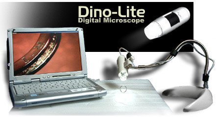
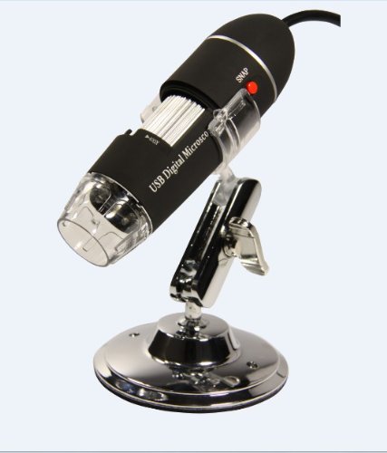
Tip 5 - Micro photography. If you can interface that camera with a microscope, you can get some honestly crazy images. Or, stack up a bunch of close-up magnification and try your hand at turning salt crystals into surreal imagery.

Tip 6 - guarnatee Photos. Ok, maybe a bit boring, but you and your friends and relatives will thank you. Take a concentrate hours and touch and photograph all of value, with a full shot or two if each item of value, accompanied by a shot of the identifying marks - manufacturer model or serial number. Then burn a Cd or Dvd and store it off site. If you have a fire or other loss, this could save the owner thousands of dollars.

Tip 7 - house formula book. Anytime those house favorites are prepared, copy down the formula and take some photos of the food. You can produce a printed or electronic cookbook of house favorites that every person will love.

Tip 8 - Stock Photography. This is a very busy store niche, but the cost of entry is low. Specialize in things you love, and you may be able to originate some wage from your stock images. Crusade for stock photography sites, and make sure you understand your possession before you post images.

Tip 9 - extra Effects. Maybe you want to specialize in high-speed images of athletes, or surrealistic collages. Try your hand at using your editing skills to put someone in a soda bottle or floating on a candy lifesaver. Often more artistic than photographic, it will test your composition, lighting and editing skills to come up with believable artificial realities.

Tip 10 - Still Life. Ahh, the bowl of fruit. Sometimes a simple object or collection, properly lit, shot and edited, is a thing of beauty. It's a great way to study light. Start with an egg on a light background, a lamp and a window and see how you can learn about lighting and composition.

Tip 11 - Computer Control. Many cameras have a Usb interface and remote operate software. You can honestly operate the camera from the computer. See if you can get it to work to your liking, and maybe even schedule some time lapse or exposure bracketing experiments.

Tip 12 - Be Like Andy. Take some images of everyday items and try to originate those neat colored backgrounds like Andy Warhol used to make. originate a 4-up print of the same image and convert the colors of each quadrant to make an tantalizing square print.

Have fun with these ideas, and let me know when you become famous!

12 Creative Photography Ideas

Related : Picking Safety Products Cabinetrack 19 inch
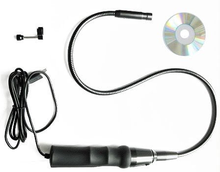