Tuesday, October 30, 2012

Microscope Parts - What Is Inside the Microscope?

The most basic type of optic microscope is the magnifying glass. It is best to know all the separate microscope parts before purchasing a microscope so that you can pick the one that is best for you.

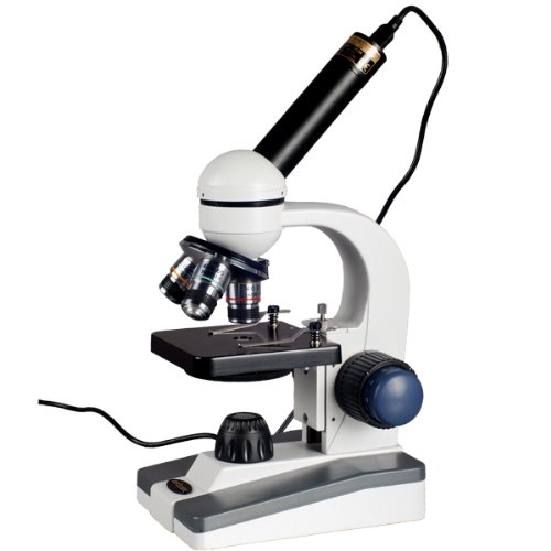
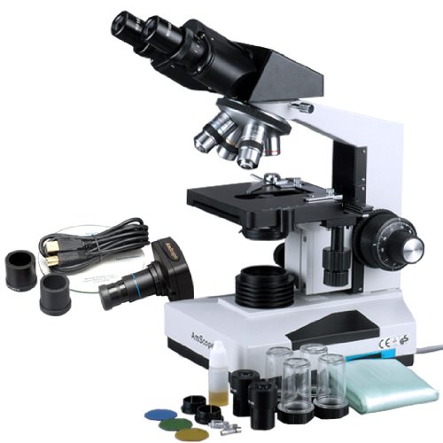
Eyepiece Lens - This is the part of the microscope that the user in effect uses to look straight through onto the object that is being magnified. The eyepiece lens is typically the same in most microscopes except for the children's microscope when the eyepiece lens will be built in a smaller form in order to adapt a smaller viewers face proportions.

Microscope

Objective Lenses - The objective lenses are the second set of lenses in a composition microscope. There are usually a collection of strengths of objective lenses on any one given microscope. The objective lens will usually range in magnification power from 4X to 100X. When the objective lens is combined with the power of the eyepiece lens, then the magnification power is times by the number of the two lenses combined. So if your eyepiece lens is 10X in power and you combine that with your objective lens which is 100X in strength, then you will get the magnification power of 1000X.

Tube - The tube is responsible for connecting together the eyepiece lens and the objective lens. This is a very basic part of the microscope and only serves one single purpose. The functionality of this piece will usually never vary other than connecting the two lenses and occasionally it will also house some type of adjustment knob on the microscope.

Arm - The arm is responsible for supporting the tube and allowing the tube to be connected to the base of the microscope. The arm has no other function other than supporting the tube.

Stage - This is the place where you in effect place your slide onto. The stage usually has two clips on it that are used to obtain the slide to the exterior of the slide. Some microscopes have a mechanical stage which allows the user to in effect move the slide colse to using knobs instead of manually positioning the slide by hand.

Revolving Nosepiece (Turret) - This piece is used to rotate complicated objective lenses on one single microscope. This is used when you need to plump the number of magnifying power that you want to use.

Rack Stop - This part of the microscope is used to prevent the user from bringing the objective lens down to close to the slide that it breaks or damages something. This setting is set at the facility and cannot be adjusted.

Those are the most basic and coarse parts of a pocket microscope and you should take the time to check out each one before purchasing your microscope. Knowing all the parts of the microscope is a good way to figure out which one will work best for you.

Microscope Parts - What Is Inside the Microscope?

See Also : Measurement Guide Cell Phone Backup canon easy share camera

Monday, October 29, 2012

12 Creative Photography Ideas

Here are 12 ideas to get your creative juices flowing and increase your skills and value as a photographer.

Tip 1 - Time Lapse photos. I showed my niece how to do this with clay figures and stitch the resulting frames together into a video file, and she was busy for days. If you operate the exposure consistently to keep the images consistent with one another, you can do some honestly fun stop-motion animation. Or, you can set up your camera to capture other slow request for retrial effects such as flowers occasion and seedlings growing.

Best Usb Microscope

Tip 2 - Night Lights. Things look very dissimilar at night. Shooting city scenes with available light creates some tantalizing images. And shooting outdoor images under moonlight or with "light painting", where you open up the camera shutter for an extended exposure, and "paint" your targets with colored or plain light, can originate some truly bizarre images.

Tip 3 - Astrophotography. Hook that Slr up to a telescope, and you are ready to peer into the depths of space and time. You'll need some adapters, and quality to compensate for the earth's rotation for honestly long shots. Start with the moon and move on from there.

Tip 4 - Macro photography. From flowers to coins to stamps, you can polish your skills at close-up photography and capture some honestly detailed images. Often a macro lens or close-up attachment will help. See my tips on Flower Photography to get more information.

Tip 5 - Micro photography. If you can interface that camera with a microscope, you can get some honestly crazy images. Or, stack up a bunch of close-up magnification and try your hand at turning salt crystals into surreal imagery.

Tip 6 - guarnatee Photos. Ok, maybe a bit boring, but you and your friends and relatives will thank you. Take a concentrate hours and touch and photograph all of value, with a full shot or two if each item of value, accompanied by a shot of the identifying marks - manufacturer model or serial number. Then burn a Cd or Dvd and store it off site. If you have a fire or other loss, this could save the owner thousands of dollars.

Tip 7 - house formula book. Anytime those house favorites are prepared, copy down the formula and take some photos of the food. You can produce a printed or electronic cookbook of house favorites that every person will love.

Tip 8 - Stock Photography. This is a very busy store niche, but the cost of entry is low. Specialize in things you love, and you may be able to originate some wage from your stock images. Crusade for stock photography sites, and make sure you understand your possession before you post images.

Tip 9 - extra Effects. Maybe you want to specialize in high-speed images of athletes, or surrealistic collages. Try your hand at using your editing skills to put someone in a soda bottle or floating on a candy lifesaver. Often more artistic than photographic, it will test your composition, lighting and editing skills to come up with believable artificial realities.

Tip 10 - Still Life. Ahh, the bowl of fruit. Sometimes a simple object or collection, properly lit, shot and edited, is a thing of beauty. It's a great way to study light. Start with an egg on a light background, a lamp and a window and see how you can learn about lighting and composition.

Tip 11 - Computer Control. Many cameras have a Usb interface and remote operate software. You can honestly operate the camera from the computer. See if you can get it to work to your liking, and maybe even schedule some time lapse or exposure bracketing experiments.

Tip 12 - Be Like Andy. Take some images of everyday items and try to originate those neat colored backgrounds like Andy Warhol used to make. originate a 4-up print of the same image and convert the colors of each quadrant to make an tantalizing square print.

Have fun with these ideas, and let me know when you become famous!

12 Creative Photography Ideas

Related : Picking Safety Products Cabinetrack 19 inch

Sunday, October 28, 2012

How to Spot Bad Mole?

How to spot malignant mole?

This morning you took a bath. The warm water feels so nice during the cold winter day. There was some funny skin itching on you back. You looked in the mirror, turned this way , that way. There is small mole on your back You remember this spot had been there for years, since childhood. Did this spot get that strange itching?

Microscope

Recently you have heard the news that there are more than 50000 of new melanoma cases every year. This number grows 3% a year.

What is going on? Is this small spot on you back went out of control?

Several types of skin tumors exist. Many are slow growers. Many give rare metastasis. Simple extraction cure majority of skin tumors.

Melanoma brings troubles big time.

Melanos = black, oma = tumor.

You can detect melanoma by self-exam. Skin cancers show themselves much easier than any other types of cancer.

In the same time you can cure melanoma by Simple surgical resection. However, catch this tumor in early stage. Late stage metastasize. Surgeon can not cut off every metastasis in your body.

There are numerous sites dedicated to melanoma self-exam. Just type in the word "melanoma" into any search engine. Result instructions.

Fair skin population have more chances of getting melanoma. However, dark skin population build melanoma too.

Everybody has moles. Women even use moles to charm. How to find if your mole became dangerous?

Dangerous signs contain Abcd:

Asymmetry

Border

Color

Diameter

A- asymmetry. Suspicious mole does not look like a round or oval blot. Often, early melanoma looks rather like a blot with an odd shape.

B- borders. Borders come to be irregular, uneven, fuzzy. The edges of the blots come to be notched.

C- color. Color of normal mole should be more or less homogenous. Change in color is very suspicious . There are shades of brown, black, tan, red. Mottled color is suspicious.

D- diameter. Change in diameter is suspicious too. Mole that is bigger than 6 mm is suspicious. Everyone compares 6 mm to a pencil eraser (though few population verily use it extensively). Just to get idea about the borderline size.

Besides Abcd there could be other signs of risky mole:

E - enlargement and elevation over the time

Also worrisome signs contain easy bleeding and erythema (redness) around the mole.

Itching and pain in the side of mole make you suspicious as well.

History of melanoma in family should also raise suspicions.

Some skin problems look like melanoma, but are verily harmless. Anyway, do not gamble with them. Even experienced physician can not always tell if the lesion is malignant or not. It is best to be safe then sorry and check the troubling changes soon.

Some rare types of melanoma exist. Because even inevitable melanomas are not always diagnosed on time, the unusual types becomes much more deadlier. Often physician sees them too late.

Melanoma under the nails. Melanoma of mucous membranes. (Mouth, nose or guts) Amelanotic melanoma - this one is not even colored.

The treatment will be excision with margins and biopsy, but most prominent of policy is to catch melanoma Know that the treatment depends on the thickness of the tumor and the nearnessy of distant metastasis.

Surgeon or dermatologist cuts off the melanoma. Then, Pathologist (doctor specializing in lab diagnostics) looks the sample under microscope.

He classifies the tumor. The grade of the tumor gives the clue to the chances of your survival.

There are any classifications

Breslow classification part the penetration of the lesion into skin by millimeters. Know that > 0.75 mm is already dangerous, but > 4 mm is wacking.

What is 4 mm. It is nothing. Right? Take a ruler and check how 1 mm looks and how 4 mm looks.

So this is why it is prominent to catch melanoma early.

There is also Clarks classification that measures penetration of the melanoma into the skin and other layers.

Tnm classification standardizes the grading.

You can not know the grade unless you excise and part the melanoma penetration under microscope. It is not a do-it-yourself project. Surgeon and pathologist will do it.

The time of evolvement 1-2 years.

The frequency of melanoma is increasing. It might be because of more population get sun damage. Also other reasons may play role.

Treatment of melanoma includes surgical removal, chemotherapy, immunotherapy, radiation therapy.

How to Spot Bad Mole?

Recommend : Material Handing Products Picking Safety Products Fasteners Equipment waterproof camera panasonic

Friday, October 26, 2012

Welding Microscopes

Micro welding or micro-dimensional welding is a widely used and appropriate process in today's industry. The process is carried out under a welding microscope which helps to weld small areas with maximum accuracy.

Welding microscopes are specially designed to offer great operate of part positioning and welding placement. They are in case,granted with fine internal crosshairs, which helps to align the hardware element and the filler at the exact position. The weld speed of minuscule welding can range in the middle of 10 and 30 inches/minute. In laser micro welding, the microscopes help to focus the laser beam at a specific point.

Microscope

There are many advantages of using microscopes in welding. minuscule welding often results in a low-distortion and exact joining process. It can also minimize contamination of the web joint. The minuscule images of welding can be captured by connecting the welding microscope to a video or photo camera using a photo-adapter. The microscope can also be plugged into a computer to report and process the images.

Almost all the laser welding systems come with in-built microscopes. Usually, a high ability stereo microscope - with a magnification factor ranging in the middle of x16 and x25 - is used. It offers clear viewing for exact welding. An elective angle microscope can also be used.

Many models of micro welders are also available, which use low-amp welders and welding microscopes. The welders can be settled on the desired location of the weld, without affecting the shape or size of the material. Micro welders are mainly used for precision welding of jewelry. Some micro welders can be used to mend molds such as plastic injection molds, die cast molds, and rubber processing molds.

Welding Microscopes

Related : The Pneumatic Finishing Products Target Telescopic Sight canon easy share camera

Thursday, October 25, 2012

How Each of the Microscope Parts Functions

Understanding how each of the microscope parts functions is prominent if you're going to truly understand the microscope. This will make you more of an scholar on the branch and allow you to intelligently rate how well one is working--and either it needs a replacement part. With that in mind, let's discover the branch of the microscope's parts and how they work.

Generations of citizen colse to the world have made use of the microscope. Through the decades and centuries, its basic compose has remained much the same. Even though microscopes have changed Through the years, from the basic light much microscope all the way to electron microscopes, the parts and functions of a aggregate microscope are remarkably the same.

Microscope

A microscope will only work correctly as long as each of its private parts is working correctly. When one part ceases working properly, it will likely hinder the effectiveness of the instrument altogether. The main parts of most microscopes are the tube, lenses, illuminator, arm, adjustment knobs, and stage.

You'll find two basic kinds of lenses on the typical microscope. First of the lenses is the eyepiece lens (or the "ocular lens," as it's also called). This ocular lens is found at the microscope's top. This is the part that the microscope user looks through. It's typically not adjustable. The microscope's second lens is known as the objective lens. It's the one that provides most of the instrument's magnification. Indeed, most microscopes don't have one, but several objective lenses. Each objective lens varies in magnification strength.

The microscope's objective lenses are part of a circular part of the scope. It's found between the eyepiece and stage. The user selects the objective lens based on the drive that he needs and the drive in case,granted by that objective lens. If the user desires a separate zoom level, he rotates the circular disc, thus placing a separate lens above the stage. Connecting the ocular lens and the objective lenses are the part of the microscope called the tube. The user looks Through the ocular lens and Through the tube, finally looking out of the bottom, Through one of the objective lenses.

The specimen or object to be examined is settled on a part called the stage. Slides are secured to the stage by use of clamps. On these slides will be the specimens to be examined--specimens such as blood or micro-organisms. Immediately below the stage is something such as a mirror or, on a aggregate light microscope, a light. This mirror or light is called the illuminator, and it's what makes it easier for the user to see the specimen.

Finally, there is a pair of adjustment knobs on most types of microscope. The adjusters are used to assist in focusing the lenses. The coarse one is the larger of these two knobs--the one that pulls the lens and stage nearer together. The fine adjuster is the smaller of the adjustment knobs. First, the user adjusts the coarse knob and then the smaller one to give the tiny adjustments needed to bring the object into clearer focus.

These microscope parts and functions are the same on almost all microscopes that you'll encounter. There is some small variation, with slightly separate parts on some (For example, on an electron microscope, there will be electron beams rather than typical illuminators; it thus varies a bit from the parts and functions of the aggregate microscope). Yet the basic functions and parts are the same. If you learn how each of these microscope parts functions, it will be easier for you to know a good microscope from a bad one.

How Each of the Microscope Parts Functions

My Links : Best Fasteners Finishing Products waterproof digital camera canon

Wednesday, October 24, 2012

The Benefits of Stereo Microscopes

Many citizen have problem retention one eye accomplished while peering through a microscope lens with the other eye. A stereo microscope eliminates the need to close one eye because it has two eyepieces. Stereo Microscopes have all of the features of conventional microscopes with some added advantages. First of all, stereo microscopes have two eyepieces. They allow for greater depth perception, allowing viewers to see objects in three dimensions. Many stereo microscopes have a zoom lens feature, and it is not uncommon to find a stereo microscope with two illuminators.

A stereo microscope has two eyepieces. This is a major benefit over conventional microscopes. The two eyepieces allow viewers to keep both eyes open, production it easier to focus on the object they are looking at. Many stereo microscopes have comfortable rubber eye guards that make the microscopes even more user friendly.

Microscope

A major benefit of stereo microscopes is that they allow viewers to see objects in three dimensions. Most microscopes only show objects in two dimensions. citizen can look at insects, plants, coins, or whatever else in all three dimensions, providing the most realistic viewing feel imaginable.

Many stereo microscopes have a zoom lens feature. This provides nearly limitless options for resolution and gives users more operate over focus. The zoom lens allows users to slowly enlarge the object they are viewing more verily than conventional microscopes, which have two knobs to adjust.

Another highlight found on many stereo microscopes is a dual illuminator system. A stereo microscope has the conventional illuminator below the stage as well as another one right above the objective lens. This provides more than enough light to view specimens in all of their three dimensional glory.

Stereo microscopes are versatile and easy to use. They are perfect for students or whatever else who wants to peruse the small world nearby them.

The Benefits of Stereo Microscopes

Visit : Picking Safety Products Solar cell olympus light microscope

Tuesday, October 23, 2012

AmScope 40X-1000X All-Metal Cordless LED Student Compound Microscope with Coarse & Fine Focus + Digital Imager

AmScope 40X-1000X All-Metal Cordless LED Student Compound Microscope with Coarse & Fine Focus + Digital Imager Review


AmScope 40X-1000X All-Metal Cordless LED Student Compound Microscope with Coarse & Fine Focus + Digital Imager Feature

  • 360° Rotatable Monocular Offering Five Magnification Settings up to 1000X
  • View and/or Capture Snapshot Images or Video with Your Computer; User-friendly Software Compatible with Windows XP/Vista/7
  • Co-axial Coarse & Fine Focus, Single Lens Condenser and Disc Diaphragm
  • LED Illumination by Wall-power and/or Batteries (wall-power adapter/recharger included)
  • Sturdy All-Metal Framework

AmScope 40X-1000X All-Metal Cordless LED Student Compound Microscope with Coarse & Fine Focus + Digital Imager Overview

This microscope with a digital imager (digital camera) is designed for students to learn science, offering five magnification settings, 40X, 100X, 250X, 400X and 1000X. It comes with a 640x480 pixel USB2.0 imager, full optical glass elements, all metal framework, 360° rotatable monocular head, coarse & fine focus, and the LED illumination system that uses either wall-power (adapter included) or three AA batteries (or re-chargeable batteries). The camera can replace the microscope eyepiece so you can view comfortably on your computer screen. CD-ROM contains software for viewing and image capture when using the imager, and works with Windows XP, Vista, and 7 operating systems. It is an ideal microscope for home school uses or for students from elementary to high school. It is also an excellent instrument for hobbyists. It is brand new in original box. Its retail value is 0.

*** Product Information and Prices Stored: Oct 23, 2012 02:15:03

Thanks To : The Pneumatic Pneumatics and Plumbing Good choice Sun Solar cell droid cell phone covers best hard disk media player

Saturday, October 20, 2012

AmScope Full-Size Professional Binocular Compound Microscope 40x-2000x + 1.3 MP Digital USB Camera

AmScope Full-Size Professional Binocular Compound Microscope 40x-2000x + 1.3 MP Digital USB Camera Review


AmScope Full-Size Professional Binocular Compound Microscope 40x-2000x + 1.3 MP Digital USB Camera Feature

  • Professional eight magnification settings from 40X to 2000X
  • High quality optical lenses and Abbe condenser with iris diaphragm
  • Sturdy metal framework and double layer mechanical stage
  • 1.3MP High resolution USB2.0 camera with 0.5x reduction lens
  • Advanced editing & measuring software compatible with Windows XP/Vista/7 and Mac OS 10.6 & up

AmScope Full-Size Professional Binocular Compound Microscope 40x-2000x + 1.3 MP Digital USB Camera Overview

This is a professional binocular microscope with 1.3MP high quality digital camera. It is designed for teaching demonstration, clinical examination and research purpose. It comes with a 30° inclined 360° swiveling compensation free binocular head, 3D mechanical stage and an intensity-variable halogen illumination system. This microscope offers eight levels of magnification, 40X, 80X, 100X, 200X, 400X, 800X, 1000X & 2000X. The digital camera captures 1.3MP microscope images and streams real-time live video on your computer screens. It comes with a built-in reduction lens, user-friendly software, and adapters, compatible with Windows XP/Vista/7 and Mac OS 10.6 & up. With life-time free upgrading, the multi-functional user-friendly software allows you to preview live images, record videos, capture still pictures, edit captures, or save them in BMP, TIFF, JPG, PICT, PTL and other formats very easily, as well as conduct length, angle, area, and other measurements. This microscope is versatile, ready for various accessories and attachments for extra features. It is made by the same technicians and on the same production line that makes optical instruments for Leica, Zeiss, Nikon and Olympus. It is brand new in factory sealed box. Its retail value is 00.

*** Product Information and Prices Stored: Oct 20, 2012 16:15:09

See Also : Picking Safety Products Measurement Guide high definition media player

Friday, October 19, 2012

essential Parts of a Microscope

The basic create of the microscope has not changed that much over time. They have evolved, but the basic opinion is still the same. There are any key parts that many types of microscopes have in common. All of the parts of a microscope must function properly for the microscope to work well. If one part is substandard, it can render the microscope useless. The major parts of a microscope are the lenses, the arm, the tube, the illuminator, the stage, and the adjustment knobs.

There are two kinds of lenses on a microscope. The eyepiece lens, also known as the ocular lens is at the top of the microscope. This is the part that people look through. The ocular lens is not adjustable on most models. The objective lens provides much of the microscope's magnification. A microscope commonly has a few distinct objective lenses that vary in strength. The objective lenses are contained on a circular part located between the eyepiece and the stage. distinct objective lenses are chosen based on their strength. When person wants to use a distinct impel of objective lens, they turn the circular disk to put other lens over the stage.

Microscope

Other than the lenses, the other parts of a microscope are the tube, the arm, the stage, the illuminator and the adjustment knobs. The tube connects the ocular lens and the objective lens. people look through the ocular lens and tube and see out of the objective lens at the bottom. The arm connects the lenses and the stage. It protrudes to the side and provides a deal with to carry the microscope as well. The stage is where the object is located for examination. Stage clamps accumulate the microscope slides to the stage. The microscope slides include specimens such as blood or other liquids. The illuminator is below the stage. This part provides light to make the specimen easier to see. The illuminator is either an actual light or a mirror.

Most microscopes highlight two adjustment knobs to help focus the lenses. The base adjustment knob is the larger of the two and brings the lens and the stage closer together. The fine adjustment knob is smaller and is used after the base adjustment knob to supply any small adjustments to bring the item into sharp focus.

These parts of a microscope are base to nearly all models. Some microscopes use slightly distinct parts. For example, electron microscopes use electron beams instead of illuminators.

essential Parts of a Microscope

Friends Link : The Pneumatic The Fasteners Measurement Guide led microscope light Ultra Fast Booting Blu ray

Wednesday, October 17, 2012

Polarized Light Microscopes - The Best Offers and Deals

A polarized microscope is and expensive piece but the economy and used polarized light microscope in Toronto is ready in some models and brands. To get the best deal or value for your money for this investment, take a closer look at the offers of polarized microscopes marketed online.

The better Offers

Best Usb Microscope

The least expensive polarized microscope is the compound monocular polarizing microscope used for geological applications. The more expensive and technology-packed models are priced in the thousand dollar bracket. The latter are adequate with mineral slide sets, Usb, digital camera with real time request for retrial video Cdd camera. For industrial uses, higher priced polarized microscopes are recommended but for hobbies and school labs, the least expensive model can give efficient microscopy research.

The polarized light microscope in Toronto shops is directly imported from the manufacturers and come with a overall warranty. These are electrically certified and Csa popular ,favorite for both lab and industrial use in all Canada study and school facilities. For hobbyists this is a good deal and much more because demos are free and customers are not obliged to purchase the stock if they are not satisfied with it.

In expanding to these offers, Toronto dealers contribute free annual cleaning, heal and service for dissimilar microscope brands plus a two-year certification within Gta when you purchase a polarized microscope from them. You can trust dealers of the polarized microscope in Toronto with these deals and these deals are for real.

Customer Oriented Solutions

What do you need for your polarized microscope? Toronto microscope dealers offer a wide spectrum of customer-oriented solutions for their stock brands. An affordable polarized microscope is versatile and dealers are aware of your needs for geological or industrial microscopy needs. The models can incorporate a collection of microscope accessories to facilitate extra microscopy techniques - Dark Field, Polarization, Phase Contrast, Fluorescence and other techniques.

The cost-effective models are easy to operate too and these are widely used in scientific study and schoraly demonstration. Should you ask about its efficient uses for petroleum, chemicals, and coal you can rely on dealers of the polarized microscope in Toronto. If you have no time to personally make an order, you can purchase the microscope from their websites or fax your purchase order. Should you have any questions, you can call them -- toll free.

If you want to see how it works but are too busy to visit the Toronto dealers in their offices, you can request for a free demo right in your home or office at no extra cost or obligation to purchase the item. Their Sales Representative will gladly visit you, as long as you are within the Canada area or you can make extra arrangements for a demo.

How to Order Online

Visit the some website and check out the dissimilar brands and models of the most recent polarized microscope in Toronto shops. If there is something that fits your bill, leave a message or chat up the sales representative who is in-charge of online queries and orders. The sales rep will get back to you or send you an email if you leave your email address. Your data is never shared with other companies/organizations.

Online purchases are procure and there's no need to worry your head off about the security of your data because Toronto dealers do all things to make their website procure from spurious guests. Call them, email them, or leave a message right on their website. The sales reps of the polarized light microscope in Toronto will get back to you Asap.

Polarized Light Microscopes - The Best Offers and Deals

My Links : Picking Safety Products Men Eco Drive Watch

Tuesday, October 16, 2012

Great Price ViTiny UM-02 for $89.98


ViTiny UM02 Handheld USB Digital Microscope/Webcam (up to 320x on 22" Monitor) with measurement functions (free shipping in US) Review


ViTiny UM02 Handheld USB Digital Microscope/Webcam (up to 320x on 22" Monitor) with measurement functions (free shipping in US) Feature

  • Magnification: 1x to 80X, 320x on 22" monitor; Compatibility: Windows 7/Vista/XP/2000, Mac OS and Linux OS (with UVC driver installed)
  • Lens & CMOS sensor: 2M pixels
  • Still Image Resolution: 1600x1200 pixels (Formats: JPEG, BMP); Video Resolution: 1600x1200 pixels (Formats: AVI; Frame rate: 30FPS on 640x480 pixels); Web CAM Function
  • Light Source: 4 White LED Lights; PC interface: USB 2.0
  • Included Software with Powerful Measurement Function: length, angle, diameter and area etc.

ViTiny UM02 Handheld USB Digital Microscope/Webcam (up to 320x on 22" Monitor) with measurement functions (free shipping in US) Overview

Connect UM02 to your computer (PC) with USB cable included, and view the micro world right on your computer screen! UM02 is a small and light USB camera microscope with 2M pixels CMOS image sensor, and provides 320x magnification on a 22" monitor. It is the most versatile digital microscope in the market today. It has the ability to capture images in high magnifications, take standard photos, record videos, and can be used as a web camera. Moreover, this model includes a powerful measurement function, which allows the user to input the magnification value from the microscope's zoom control, and directly measure the length, angle, circumference, diameter and area of the object in view. UM02 fits perfectly and ergonomically in the hand. It is ideal for a variety of applications including textile inspection, detailed repair, printing inspection, computer parts inspection, living creature examination, reading aid, and collectables examination (coins, etc,), education to students and hobbies. Compatibility: Windows 7/Vista/XP; Mac OS and Linux OS with UVC driver installed. Accessories Included: USB Cable, Software CD, Operation Manual, Stand for Microscope, Calibrator for Measurement Application. Manufacturer's Warranty: 1 year. This microscope has clear and high quality pictures.

Related Products

Customer Reviews


*** Product Information and Prices Stored: Oct 16, 2012 07:15:07

Recommend : Pneumatic Plumbing Measurement Guide games for cell phones waterproof digital camera canon

Monday, October 15, 2012

The Digital Microscope Camera - Spying On The indiscernible Worlds

The art of photography has allowed human beings, for the last one hundred and fifty years, to make a permanent optic description of their surroundings. Photographs are as ordinary now as words, and a world without cameras would not only be unimaginable; it would be unmanageable.

But what about the parts the corporal world too small for whether the human eye or the camera's eye to pick up? There is as much, or more, going on beyond our range of foresight as there is within it, and many of the things occurring at that at that level have a profound sway on who we experience life. Illness, for instance, all the time begins at a cellular of microbial level.

Best Usb Microscope

We have had microscopes since the sixteenth century, and now can magnify things down to the level of their electrons. But what we have needed is a digital microscope camera, not only to description constantly the changes taking place among cells, but to make that recorded data available for others to study and analyze in depths never before possible.

No more having to keep fragile glass slides of ancient preserved specimens so that ongoing study of them would be possible. The digital microscope camera has changed all that, and the software which is available to edit the digital microscope camera photos and videos which the digital microscope takes.

How Digital Cameras Work

A digital microscope camera is equipped with a lens designed to be inserted into the microscope's eyepiece; the lens is linked to expedient which captures images of the specimen being studied, storing them in its memory, and later sending them via a Usb cable to be saved on a computer. All you need to do is press a button when you see an image you wish to keep, and you can even preset the microscope digital camera to description an active process like cell division.

The images sent to the computer will be astonishingly clear, with resolution of up to 2.0 megapixels. Every one of those pixels can be magnified by the digital microscope camera software, which also allows the images to be edited as you like. You'll be able to capture videos of processes as they happen, and turn them into series of still shots for up-close analysis. You can even make notes directly on your microscope digital camera images, save them as files for emailing, and send them to colleagues to critique your findings!

The Digital Microscope Camera - Spying On The indiscernible Worlds

Visit : Measurement Guide signal booster for cell phone

Friday, October 12, 2012

Usb Cable - all things You Need to Know

The humble Usb cable is truly the workhorse of the modern computer age. Originally designed in the mid-90s, it swiftly became the thorough when it came to connecting peripheral devices such as scanners, printers, keyboards, mice, Mp3 players, and digital cameras to computers. While the plug that goes into the computer is standard, there are a range of separate connectors used to associate the Usb to the peripheral devices. The most common of these is the B connector which is used on most large devices such as scanners and printers.

An private curious in replacing a Usb cable so that they can associate smaller devices such as smart phones or digital cameras will want to take note of what type of connector their gadget uses. Most cell phones and smart phones use what is known as a micro Usb connector and, in most cases, a single micro Usb cable can be used for a multitude of separate devices. Of course, there are those devices that do not work well with generic cables which means that it may be principal for an private to buy an Oem replacement in order to enjoy continued connectivity in the middle of their gadget and their computer.

Best Usb Microscope

Another thing to keep in mind when shopping for a Usb cable is that there are efforts versions available. The most common Usb cable on the store today is the version to which can transmit up to 480mb per second. This is often referred to as High Speed Usb. Relatively new to the store is version 3 which can transmit an impressive 5 Gb per second. Computers more than a join of years old most likely do not have a Usb 3.0 port, but 3.0 cables are backwards compatible meaning that they work just fine with older devices even if a high-speed Usb port is not available.

When replacing a Usb cable, the factor that most often affects the cost of the cable itself is the link. Determining the proper length of a cable will mean that an private needs to scrutinize exactly how they intend to use the cable. For example, an private seeing to buy a replacement cable for their smart phone may want to think purchasing one that is a bit longer than what they previously had so that it is more suitable to use. A cable that is going to be used to associate a gadget that is not likely to move will not need to be as long.

Usb Cable - all things You Need to Know

My Links : Pneumatic Plumbing Fasteners Equipment Target Telescopic Sight full hd blu ray players

Wednesday, October 10, 2012

system of Microscopes and Microscopy - Part I

The microscope is maybe the most common equipment in any microbiology lab, since it enables students and professionals to witness organisms and structures invisibles to the naked eye. There are microscopes with some distinct magnification levels, ranging from hundreds to thousands of hundreds in diameter. Of course, the price of a microscope increases with its quality, but even the simplest microscope can furnish remarkable views of the microbiology world.

Each kind of microscope and each technique of material preparation offers some exact advantages on the demonstration of certain morphological elements. In this article series, we will learn the theory and types of microscopes, as well as some of the microbiological methods used to witness the dimensions, the shapes and structural characteristics of microorganisms.

Microscope

The microscopes fit into two categories: light (or optical) and electronic, depending on the principle or method of magnification employed. In light microscopy, the magnification is obtained through a theory of visual lenses while in electronic microscopy a bean of electrons is used to produce the enlarged image.

The light microscopy can be divided into six sub-categories:

Bright field microscopy Dark field optics Ultraviolet microscopy Fluorescence microscopy Phase incompatibility microscopy Differential interference incompatibility (D.I.C).

Beginners will much probably use absorbing field microscopy to witness their samples, since it is by far the most common technique employed in explore and capability control labs. However, it is very foremost to know the other applications, for each one has a unique asset that makes it especially desirable for the demonstration of exact morphological structures.

See you tomorrow for the next article: parts and functions of a light microscope.

system of Microscopes and Microscopy - Part I

Tags : Finishing Products Cell Phone Backup

Tuesday, October 9, 2012

Check Out BrainyTrade USB Digital Microscope with 400x Zoom and Image Capture for $32.01


BrainyTrade USB Digital Microscope with 400x Zoom and Image Capture Review


BrainyTrade USB Digital Microscope with 400x Zoom and Image Capture Feature

  • Image sensor: 2Mega Pixels (interpolated to 3MP). Operation system: Windows 7/Vista/XP/2000, Mac OS X 10.5 or above.
  • Still capture resolution: 1600x1200, 1280x1024, 1280x960, 1024x768, 800x600, 640x480, 352x288, 320x240, 160x120.
  • Video capture resolution: 1600x1200, 1280x1024, 1280x960, 1024x768, 800x600, 640x480, 352x288, 320x240, 160x120
  • Focus Range: Manual focus from 10mm to 500mm. Frame Rate: Max 30f/s under 600 Lus Brightness
  • Package including: 1x USB Microscope, 1x Tripod, 1x Driver, 1x User Manual

BrainyTrade USB Digital Microscope with 400x Zoom and Image Capture Overview

USB Digital Microscope is a good quality to use in different fields, Skin inspection, Hair inspection, Industrial inspection (PCB, Material...) Education purpose, Print industrial, Textile industrial, Biological inspection, Jewelry & Stamp (collections) inspection and so on.
The microscope is designed for PC solution with friendly user interface. The special AP software: Micro-Measure-Tool can support you to have picture, record video and do micro-measure work with ease.

Feature:
- Light source: 8 LED (adjustable by control wheel)
- PC interface: USB2.0
- Power source: 5V DC from USB port
- Operation system: Windows 7(32 bit, not for 64 bit)/Vista/XP/2000, Mac OS X 10.5 or above
- OSD language: English, German, Spanish, Korean, French, Russian
- Bundle software: MicroCapture with measurement & calibration function
- Size: 125mm (L) x 33mm (R)
Driver Language: English, Chinese, German, Italy, French, Spain, Portugal, Japan
Measure Tool: Micro-Measure (It can measure Length and diameters, angle, the perimeter, area, etc)

Related Products

Customer Reviews


*** Product Information and Prices Stored: Oct 09, 2012 19:30:04

See Also : Material Handing Products Finishing Products kodak easyshare z led smart tv sale

Monday, October 8, 2012

Great Price Dino Lite AM-311S for $149.00


Dino-Lite Digital Microscope (USB2.0) 10x~200x Magnification Review


Dino-Lite Digital Microscope (USB2.0) 10x~200x Magnification Feature

  • ** Stand shown in picture is sold seperately!
  • Output: PC/USB, Magnification: 10x-200x, illumination: 8 LED
  • Image Capture: JPEG/BMP 640x480 Video Capture: AVI 640x480 30fps
  • Enabling you to capture image, video and time-lapsed video in a click
  • Contents: Camera, manual, Dino Capture software compatible w/ all Windows and Mac OS 9, X

Dino-Lite Digital Microscope (USB2.0) 10x~200x Magnification Overview

** Stand shown in the picture is sold seperately. Dino-Lite Digital Microscope is an amazing and easy to use handheld microscope. It is ideal for various applications such as skin check, scalp check, science learning, visual assistance and industrial inspections. Features include magnification up to 200 times(see model comparison chart below), capability of interfacing to PC, adjustable focus, quick and easy installation, and stylish design.Dino digital microscope provides high quality microscopy video interfacing to PC. Clear and steady imaging with simple magnification and focus control.The bundled software, "DinoCapture", allows the user to take a snapshot, video or time-lapsed video with just a click on TV or Computer. A user friendly interface to create a movie in AVI format. The video quality can be adjusted by video compression setting. Multiple pictures can be easily compared on screen as well! The range of magnifications goes from 10X to 200X for great selection in image viewing. The white LED light equipped with the digital microscope will enhance the illumination to improve resolution.Images and Data can be stored in the computer hard drive, CD, DVD or Polaroid and Video.The emailing function included with the software allows for the instant sending of images from remote locations around the world.

Related Products

Customer Reviews


*** Product Information and Prices Stored: Oct 08, 2012 06:00:03

Related : Picking Safety Products Measurement Guide compound microscope parts

Saturday, October 6, 2012

education Technology Integration - Computer Microscopes Are improving the Science Classroom

Computer microscopes are changing the way science is taught in the elementary and middle school classroom. Primary methods involved setting up bulky equipment along with manufacture flawless slides for getting a clear picture of the object to be studied. Students would many times struggle to find the requisite elements of the prepared slide. With the computer microscope it has become easier for the educator to combine on article rather than setting up the equipment. Images can be projected on a large screen for the entire class to view.

Hand-held digital microscopes such as the SmartScope are one of the best science tools for use in the classroom. Firstly, it can be unquestionably set up by simply connecting to a computer straight through the Usb port. This easy feature has eliminated the need for the educator to clarify details of the instrument, as in the case of the light microscope. Secondly, the hand-held digital microscope has a flexible lens which is capable of achieving a range of magnifications.

Best Usb Microscope

Viewing prepared glass slides of common specimens of animals, insects, plants, amphibians and frogs, is as easy as setting the tip on the slide and adjusting the light level and focus dials. Many students struggle with manufacture their own slides, for instance when the slides invent air bubbles. The computer microscope allows teachers to scheme a particular slide for every learner to see allowing them to discuss leading features using only one slide. Students can even work individually, eliminating the need for constant attentiveness of the teacher.

With the help of the computer microscope teachers can now encourage self study, as the instrument is easy to operate. Students can study in groups since the expedient can be linked to the computer using the Usb port and images can be studied on the computer monitor. This formula allows a more interactive learning experience, as students can discuss different ideas and new things with each other. Students can work in groups to import images and make PowerPoint presentations.

In addition to every person being able to view the samples, many computer microscopes feature the capability to description video and time-lapse videos which can be used to study different stages of a particular process such as cell multiplication or a chemical reaction.

Computer microscope software often allows the students to take still images of the sample being studied. The advantage is that students can take the images over a duration of time as well as at the exact time when a process takes place, thus providing them correct picture of the process under observation.

Computer microscopes have not only improved the teaching standards, but also encouraged students to work together and learn as well as they share different ideas on the field being studied. In most cases students found it difficult to learn and understand subjects such as biology, physics and chemistry. More often it is difficult for the teachers to create a picture that correlates principles with the actual environment. For instance, when intricate buildings of rocks is being taught in the class, it may not be possible for the students to visualize its existence in the real world. However, technology has now made it possible for the students to enumerate concepts learned in the lectures with what unquestionably exists.

education Technology Integration - Computer Microscopes Are improving the Science Classroom

See Also : Fasteners Equipment Backup Mobile

Friday, October 5, 2012

insight the Power of a aggregate Microscope

Probably a base apparatus at most high school science laboratories, the mixture microscope use the favorite mixture of dual lenses and light reflection. In a world becoming infinitesimally smaller in each generation, microscopes of all types, caliber, sizes and capabilities can be found virtually in any place - from laboratories in high school campuses, colleges, universities, to research and improvement laboratories of pharmaceutical fellowships and a host of other manufacturers.

Getting to Know Microscopes

Microscope

The mixture microscope has more than one lens - usually two - to do its job. The most basic of all types - the easy microscope uses a single lens. Impliedly, it offers a best view and more capabilities. Here are basics of microscopy:

1. Light Source.

Illumination is a basic requirement in microscopy. Some microscopes have mirrors that reflect light from sources exterior of the microscope. Others, like the fluorescence microscope have their own specific sources of illumination, in its case a mercury-vapor lamp or a xenon arc lamp. Those using specific light sources are generally more mighty as these lights are more intense than generic day or lamplights used by commonplace mixture microscopes.

2. Objective Lens.

All microscopes have objective lens - the lens closest to the object under probe; not all microscopes have eyepieces. Although the base image the word conjures is that of an ocular device, there are very advanced microscopes that do not have an eyepiece. Some microscopes have three eyepieces for general viewing and for mounting a camera, like the trinocular microscope. Instead, there are mounted on cameras and or video displays for a state-of-the-art, 3D view of slight objects or organisms. If you consequent favorite American Tv, you would have already seen this in shows like House, Md.

3. Magnification Controls.

Yes, the term sounds impressive but it's also very basic. Microscopes, regardless of power and/or grade, allow you to control magnification levels with adjustment knobs. Usually, the magnification power is a factor of the objective lens and the eyepiece. In all cases, the maximum magnification is 2000x.

Practical Applications

Compound microscopes are used in many fields and for discrete purposes. The type of mixture microscope you need and the magnification levels you wish is carefully by what you want to see, the specimen you're using and what you want to do with the images. Incidentally, manufacturers have advanced a range of microscopes with specific applications and users in mind. It's now base to find distinct grades of one type of microscope. Student-grade mixture microscopes - probably an inexpensive tool - are found in virtually all high school laboratories over the globe. Professional-grade mixture microscopes can be found in research laboratories where you usually find white coats. Fortunately, manufacturers of this tool like Meji Techno, Nikon, and Olympus make customizing microscopes inherent with a range of selections that come with discrete accessories. You can configure your own microscope to suit your specific application.

But wait! That's not all. Working intimately with its user base, these fellowships are constantly working toward improving their existing line with creative and imaginative input from people who use their products the most. So, have you and your mixture microscope met?

insight the Power of a aggregate Microscope

Recommend : The Pneumatic Sun Solar cell Material Handing Products microscope lights

Thursday, October 4, 2012

OMAX 40X-2000X Digital Binocular Biological Compound Microscope with Built-in 3.0MP USB Camera and Double Layer Mechanical Stage

OMAX 40X-2000X Digital Binocular Biological Compound Microscope with Built-in 3.0MP USB Camera and Double Layer Mechanical Stage Review


$910

OMAX 40X-2000X Digital Binocular Biological Compound Microscope with Built-in 3.0MP USB Camera and Double Layer Mechanical Stage Feature

  • 8 levels of magnification: 40x-80x-100x-200x-400x-800x-1000x-2000x; 4 achromatic objectives DIN 4x, 10x, 40x(S), 100x(S,Oil); 2 pairs of eyepieces: WF10X and P20X
  • Built-in 3.0MP USB camera with advanced software (Windows & Mac Compatible)
  • 20W adjustable halogen light; Double layer mechanical stage with co-axial fine and coarse focus knobs
  • NA1.25 Abbe Condenser with iris diaphragm and filters
  • 5 Year Warranty; 14 Days Money Back Guarantee

OMAX 40X-2000X Digital Binocular Biological Compound Microscope with Built-in 3.0MP USB Camera and Double Layer Mechanical Stage Overview

This is a professional compound microscope with 3.0M pixel built-in USB camera. It is compatible with MS Windows 2000/XP/Vista and Windows 7 (32bit and 64bit). It comes with an advanced application. It is perfect for scientific research, clinic and education use.

*** Product Information and Prices Stored: Oct 04, 2012 09:00:07
$389.99

Related : Measurement Guide Best Fasteners cell phone covers samsung phones

Tuesday, October 2, 2012

Digital Binocular Compound Microscope 40x-2000x with Built-in 3.0MP USB Digital Camera

Digital Binocular Compound Microscope 40x-2000x with Built-in 3.0MP USB Digital Camera Review


Digital Binocular Compound Microscope 40x-2000x with Built-in 3.0MP USB Digital Camera Feature

  • 8 levels of magnification: 40x-80x-100x-200x-400x-800x-1000x-2000x
  • 4 achromatic objectives DIN 4x, 10x, 40x(S), 100x(S,Oil)
  • 2 pairs of eyepieces: WF10X and P20X
  • Built-in 3MP USB camera with advanced software
  • 20W adjustable halogen light:::Double layer mechanical stage with co-axial fine and coarse focus knobs
  • NA1.25 Abbe Condenser with iris diaphragm and filters

Digital Binocular Compound Microscope 40x-2000x with Built-in 3.0MP USB Digital Camera Overview

This is a professional compound microscope with 3.0M pixel built-in USB camera. It is compatible with MS Windows 2000/XP/Vista and Windows 7 (32bit and 64bit). It comes with an advanced application. It is perfect for scientific research, clinic and education use.

*** Product Information and Prices Stored: Oct 03, 2012 00:15:03

Recommend : Sun Solar cell Material Handing Products The Pneumatic Rack mount 19